| Journal of Clinical Question. 2025; 2(3): e76 https://doi.org/10.69854/jcq.2025.0019 Advance access publication date 30 June 2025 |
 |
Case Report
Twin Block Appliance for Postoperative Treatment of Intracapsular Condylar Fracture in a Child: A Case Report
Xue Meng 1, Hao Chen1,2, Weixian Liu1, Lijun Fang1,*
1Department of Oral and Maxillofacial Surgery, Shengjing Hospital of China Medical University, Shenyang Liaoning, China.
2Department of Oral and Maxillofacial Surgery, The First Affiliated Hospital of Zhengzhou University, Zhengzhou Henan, China.
*Corresponding Author: email: fanglj@sj-hospital.org
Submitted: May 19, 2025 Accepted: June 30, 2025
Clinical Question Box
Is the Twin Block appliance with headgear effective in treating pediatric intracapsular condylar fractures with displacement?
The Twin Block appliance combined with headgear successfully rehabilitated a child with a displaced intracapsular condylar fracture. It corrected mandibular retrusion, restored occlusion, and promoted joint recovery without adversely affecting developing tooth germs. After 4 months, the patient achieved normal occlusion and full mouth opening, which supports its use as a safe and practical option in similar pediatric cases.
Abstract
Background: Pediatric intracapsular condylar fractures are common and often challenging to manage, particularly when accompanied by displacement and tooth loss. Case Presentation: An 8-year-old girl presented with different types of fractures on both sides, along with tooth loss. A Twin Block functional appliance was employed following surgical treatment. After treatment and 10 months of follow-up, the bilateral condyles and articular fossae had healed well, mouth opening exhibited a normal pattern, and lateral movement of the mandible was good. Conclusion: The Twin Block appliance appears to be a feasible option for the postoperative management of pediatric intracapsular condylar fractures, particularly in cases involving mandibular molar loss. Its successful application in bilateral condylar fractures offers a novel treatment option.
Keywords: Twin Block appliance, condylar intracapsular fracture, pediatrics, case report
Introduction
Pediatric intracapsular condylar fractures (ICFs) account for 20%–50% of all mandibular fractures and continue to be a topic of debate regarding optimal treatment.1 Some experts support conservative management, citing the condyle’s capacity for growth and remodeling.2,3 Others advocate for early open reduction to reduce the risk of complications such as joint ankylosis.4-6 However, certain studies have reported no significant long-term differences or complications between these two approaches.7-9 Treatment decisions are also guided by fracture classification systems, with the He classification widely used in China.10 In this system, Type A fractures are more likely to progress to ankylosis and may warrant early surgical intervention, while Types B, C, and M are generally treated conservatively.11
Cases involving bilateral ICF of different types accompanied by tooth loss are rare. The Twin Block appliance, widely used for early skeletal Class II malocclusion, is recognized for its effectiveness, ease of use, and high patient compliance.12,13 Given the limited postoperative treatment options in such complex cases, this report presents a novel clinical approach using the Twin Block appliance for postoperative management, offering a new therapeutic perspective.
Case Presentation
Initial Presentation
An 8-year-old girl was admitted to the pediatric intensive care unit with a craniocerebral injury 5 hours after falling from a building, with no prior medical history. Lip and chin wounds were debrided and sutured in the emergency department. Clinical examination revealed mandibular retrusion, a Class II molar relationship, a Grade III deep overbite, a 7-mm overjet, exfoliated deciduous molars with gingival tears, crown fractures of permanent molars, and absence of occlusal contact. Mouth opening was approximately 15 mm with a normal opening pattern, although tenderness was noted in both temporomandibular joints (TMJs) (Fig. 1A and B). CT imaging revealed bilateral ICFs, Type A on the left and Type B on the right, along with a non-displaced left mandibular fracture (Fig. 2A and B). Because of multiple injuries, including a leg fracture requiring implantation, the condylar fractures were initially left untreated, and magnetic resonance imaging was not feasible. Seventeen days later, owing to reduced mouth opening (10 mm) and absent joint mobility, open surgery was performed.
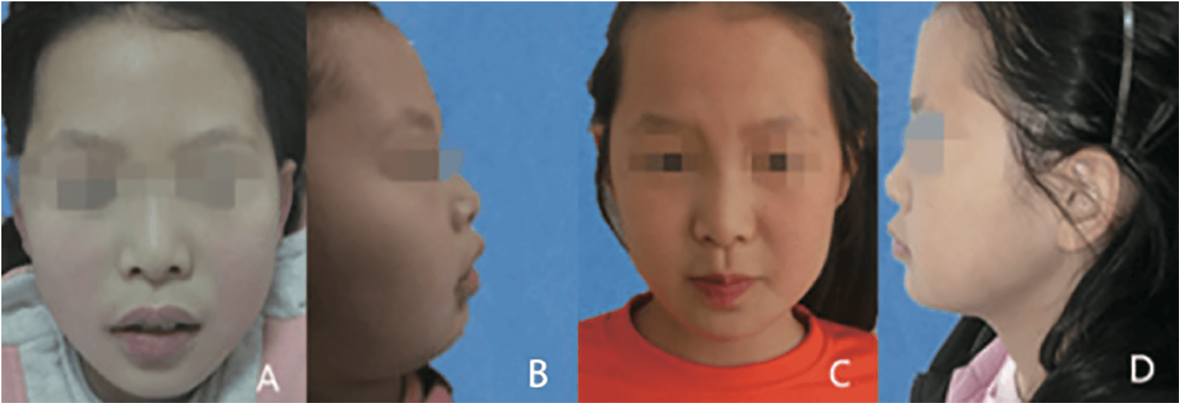
Figure 1. Frontal and lateral facial photographs before and after treatment. (A and B) Before treatment, the patient’s mandible had shrunk back, with a deep overbite facial profile and slight mandibular deviation. (C and D) After treatment, the child’s face was symmetrical, the mandibular retrusion had significantly improved, and the lower third of the face had lengthened.
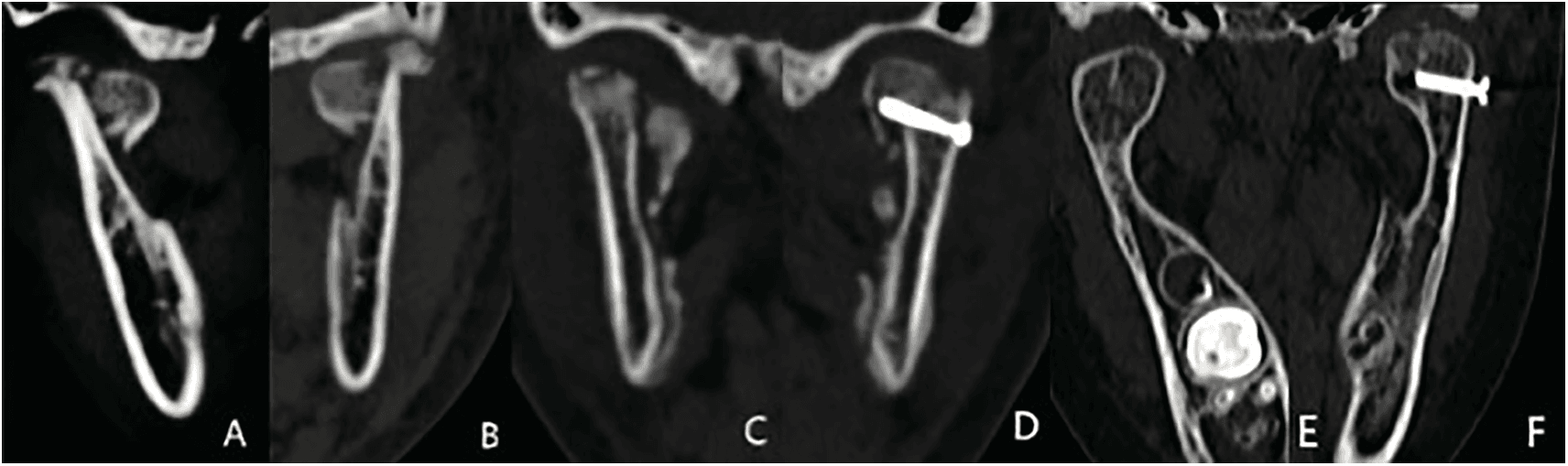
Figure 2. Mandibular computed tomography images before and after treatment. (A and B) CT images of the mandible following the traumatic injury, showing bilateral intracapsular condylar fractures, with the left condyle adhered to the lateral side of the articular fossa. (C and D) Changes in the left condyle after surgery, with the fractured end significantly reduced. The fracture fragment of the left condyle has shifted with absorption. (E and F) The condyle was in satisfactory shape after 10 months, with visible joint space.
Treatment Process
A modified pretragal approach was utilized to protect the facial nerves and auriculotemporal neurovascular bundles.14 The left joint capsule was exposed, the fracture site identified, and the callus removed to realign the fracture. The anteromedial fractured condyle was reduced with preservation of the lateral pterygoid muscle and condylar cartilage, and the occlusal relationship was confirmed. Fixation was achieved using a lag screw. Intraoperatively, mouth opening was assessed; removal of the lateral callus from the left condyle improved opening, while the right condyle was not surgically treated (Fig. 2C and D). The left joint disc was repositioned, the capsule closed, and layered suturing completed. Postoperatively, the patient received liquid food and began mouth-opening exercises.
One week postoperatively, a dental impression was taken to design an ideal occlusal position, and the mouth opening was 3.5 cm when the Twin Block appliance was fabricated. Owing to the patient’s vertical facial pattern, an end-to-end anterior bite was adopted, with a 2 mm increase in occlusion and a 1–2 mm increase in intermolar space to reduce overbite and overjet. The maxillary occlusal plate covered the 16, 14, 24, 65, and 26 plane, forming a 45° mesial-to-distal guiding incline (Fig. 3). An arrow clasp was placed on the maxillary first molars, an interdental clasp between the premolars, and a transpalatal arch connected the first molars. A labial bow retracted the maxillary anterior teeth. The mandibular molars were exfoliated owing to trauma, and the permanent premolars had not erupted; thus, an interdental clasp was placed between the 12, 11, 21, and 22 anterior teeth. Glass-ionomer cement extended the clinical crown height of 16 and 26 for occlusal support. The headgear was fitted for mandibular traction.

Figure 3. Twin block appliance retention and occlusal outcome. (A and B) The appliance demonstrated good retention during treatment; the bilateral molars were missing. (C and D) After treatment, the occlusal relationship was satisfactory, with significant improvements in both deep overbite and deep overjet.
Appliance Adjustment and Postoperative Follow-up
After the appliance was fitted, retention was checked, and the child was instructed to wear it 24 hours a day. One week later, a follow-up was performed to relieve pain, followed by visits every 4–6 weeks to assess sagittal and occlusal relationships, adjust the maxillary and mandibular pads, evaluate traction, and monitor the bilateral TMJs with panoramic radiographs and mandibular CT (Figs. 2E, F and 4). Records included facial nerve function, TMJ status, facial development, and mouth opening (Fig. 1C, D).
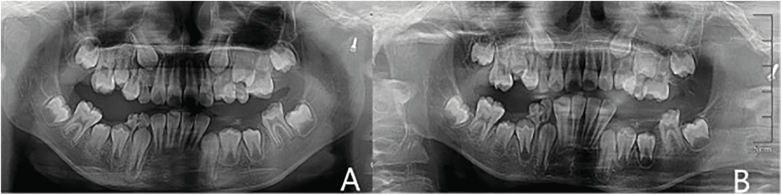
Figure 4. Panoramic radiographs showing mandibular root healing and orthodontic potential A: Root tip trauma of the mandibular anterior teeth, missing deciduous molars, crown defect of 36, and left condylar implant. B: Four months after treatment, the shadow at the root tips of the mandibular anterior teeth had disappeared, indicating that orthodontic force can be achieved by relying on the mandibular anterior teeth.
Postoperatively, mandibular retrusion improved. The left eyelid, initially slow to close, recovered within 1 month. CT imaging showed the left condyle was significantly reduced in the articular fossa with preserved disc space; the right condyle was in place with an acceptable shape. The patient adapted to the Twin Block appliance within 1 week, maintaining normal chewing and speech. At 2 months, anterior overjet was 4 mm, deep overbite was Ⅱ°, the lower face had lengthened, and the mentolabial sulcus became shallower (Fig. 1E, F). By 4 months, mandibular molar occlusion was established, overbite normalized, overjet was approximately 3 mm, and the appliance was removed without adverse events. Mouth opening reached 37 mm with a normal pattern and good lateral movement. At 10 months, CT imaging confirmed satisfactorily healed bilateral condyles and fossae with normal joint spaces and morphology (Fig. 2E, F), and the patient expressed satisfaction with the outcome.
Discussion
This case presents a pediatric patient with bilateral ICF of different types, mandibular molar loss, and limited mouth opening, treated with open reduction on one side followed by functional rehabilitation using a Twin Block appliance combined with headgear. This orthodontic postoperative approach is novel in managing pediatric condylar fractures with tooth loss, offering effective restoration of occlusion, mandibular function, and joint mobility. This case highlights a promising, noninvasive adjunct for postoperative rehabilitation in growing patients with complex mandibular injuries.
The treatment of ICF in children requires an individualized approach to ensure functional recovery while minimizing long-term complications such as joint ankylosis, mandibular asymmetry, and growth disturbances. In the present case, limited mouth opening necessitated open surgery. Postoperative rehabilitation plays a critical role and typically includes occlusal guidance and the use of functional appliances.1 An occlusal pad was applied postoperatively to raise the bite and lower the mandibular ramus, facilitating restoration of condylar morphology, repositioning within the articular fossa, fracture healing, and correction of trauma-induced occlusal and neuromuscular dysfunction.15 Standard occlusal pad therapy relies on accurate molar occlusion, bilateral molar elevation, and anterior teeth traction. Common traction methods include screws, orthodontic brackets, and intermaxillary elastics. However, these are often unsuitable for children with extensive occlusal trauma, anterior tooth injury, or missing molars, and the use of traction screws in mixed dentition poses a risk of damaging developing permanent tooth germs.
Although the Herbst appliance has shown promising results in treating pediatric condylar fractures, it requires molar anchorage and is therefore unsuitable in cases with molar loss.16 In this case, a Twin Block appliance was selected. This appliance comprises separate upper and lower acrylic plates with bite blocks that position the mandible forward. It facilitates skeletal and dental correction during growth by guiding mandibular development and improving the sagittal jaw relationship.17 Compared with standard occlusal pads, the Twin Block combined with headgear offers several advantages: ease of removal and cleaning, protection of developing tooth germs, suitability in cases with molar loss, early initiation of functional training, and stable occlusal outcomes.18 Traditional Twin Block treatment is divided into two phases: an active phase lasting approximately 6–12 months, and a maintenance phase of 4–6 months.19 In this case, the patient wore the appliance for 4 months and achieved a favorable treatment outcome. However, its limitations include potential discomfort and the need for consistent patient cooperation, making it inappropriate for patients with severe brain injury or cognitive impairment.20
A literature search using the keywords “Intracapsular Condylar Fracture” and “Twin Block” yielded no relevant results in PubMed. The primary limitation of this case report is that it is based on a single case with limited follow-up.
Conclusion
The Twin Block appliance is an effective postoperative option for pediatric ICFs, particularly in cases involving mandibular molar loss. It supports occlusal recovery and joint remodeling while minimizing risks to developing tooth germs, making it ideally suited for growing patients.
Acknowledgment
We appreciate the patient’s cooperation in data collection.
Funding Source
This study was funded by the Doctoral Program Foundation of Institutions of Higher Education of China (No. 20132104110012) and was partially supported by the Japan–China Sasakawa Medical Fellowship.
Author Contributions
X.M. was responsible for data curation, data interpretation, and drafting of the original manuscript. H.C., W.L., and L.F. substantially contributed to revising the manuscript draft. All authors have read and approved the manuscript and agree with its content and data.
Data Availability
The datasets used in the current study can be obtained from the corresponding author upon reasonable request.
Ethical Statement
The patient’s guardians provided written informed consent for the publication of this report and accompanying images.
Conflict of Interest
The authors report no conflicts of interest in this work.
Supplemental Information
Supplemental information for this article can be found online at https://sup.jclinque.com/api/articles/76/download-suppl.
References
[1] Bottini GB, Roccia F, Sobrero F. Management of pediatric mandibular condyle fractures: a literature review. J Clin Med. November 17, 2024;13(22):6921. doi:10.3390/jcm13226921.
[2] Vanpoecke J, Dubron K, Politis C. Condylar fractures: an argument for conservative treatment. Craniomaxillofac Trauma Reconstr. March 2020;13(1):23–31. doi:10.1177/1943387520902881.
[3] Monarchi G, Catarzi L, Paglianiti M, Valassina D, Balercia P, Consorti G. A comparative analysis of surgical and conservative management in intra-articular condylar fractures: a retrospective study. Surgeries. 2024;5(4):1033–1042. doi:10.3390/surgeries5040083.
[4] Akkoc MF, Bulbuloglu S. The treatment perspective of pediatric condyle fractures and long-term outcomes. Cureus. October 2022;14(10):e30111. doi:10.7759/cureus.30111.
[5] Karan A, Kedarnath NS, Reddy GS, et al. Condylar fractures: surgical versus conservative management. Ann Maxillofac Surg. January–June 2019;9(1):15–22. doi:10.4103/ams.ams_157_17.
[6] Lai BR, Liao HT. The comparison of functional outcomes in patients with unilateral or bilateral intracapsular mandibular condylar fractures after closed or open treatment: a 10-year retrospective study. Ann Plast Surg. April 1, 2023;90(1 Suppl 1):S19–s25. doi:10.1097/sap.0000000000003346.
[7] Ohayon C, Perelman A, Katz Biton A, et al. Conservative vs. surgical management of condylar fractures in pediatric populations: complications and factors for consideration. Children (Basel). March 3, 2025;12(3):323. doi:10.3390/children12030323.
[8] Minervini G, Franco R, Marrapodi MM, Di Blasio M, Isola G, Cicciù M. Conservative treatment of temporomandibular joint condylar fractures: a systematic review conducted according to PRISMA guidelines and the Cochrane handbook for systematic reviews of interventions. J Oral Rehabil. September 2023;50(9):886–893. doi:10.1111/joor.13497.
[9] Dai J, Gong C, Diarra D, Li Z. Outcomes of removing the fracture fragments in the treatment of intracapsular condylar fractures in children. Med Oral Patol Oral Cir Bucal. February 2025. doi:10.4317/medoral.27079.
[10] He D, Yang C, Chen M, Jiang B, Wang B. Intracapsular condylar fracture of the mandible: our classification and open treatment experience. J Oral Maxillofac Surg. August 2009;67(8):1672–1679. doi:10.1016/j.joms.2009.02.012.
[11] Boffano P, Benech R, Gallesio C, Arcuri F, Benech A. Current opinions on surgical treatment of fractures of the condylar head. Craniomaxillofac Trauma Reconstr. June 2014;7(2):92–100. doi:10.1055/s-0034-1371772.
[12] Rezky Oktaviyani R, Harun A, Wesley K, et al. Myobrace versus twin block in the treatment of class II malocclusion in Children: a systematic review. Saudi Dent J. May 2024;36(5):661–664. doi:10.1016/j.sdentj.2024.03.006.
[13] Tüfekçi E, Allen SB, Best AM, Lindauer SJ. Current trends in headgear use for the treatment of Class II malocclusions. Angle Orthod. July 2016;86(4):584–589. doi:10.2319/041315-242.1.
[14] Li H, Zhang G, Cui J, Liu W, Dilxat D, Liu L. A modified preauricular approach for treating intracapsular condylar fractures to prevent facial nerve injury: the supratemporalis approach. J Oral Maxillofac Surg. May 2016;74(5):1013–1022. doi:10.1016/j.joms.2015.12.013.
[15] Yang C, Zhang S, Zhang Y. Three-dimensional-printed splint for use in pediatric mandibular fracture. J Craniofac Surg. March–April 1, 2023;34(2):e186–e187. doi:10.1097/scs.0000000000008984.
[16] Al-Dboush R, Soltan R, Rao J, El-Bialy T. Skeletal and dental effects of Herbst appliance anchored with temporary anchorage devices: a systematic review with meta-analysis. Orthod Craniofac Res. February 2022;25(1):31–48. doi:10.1111/ocr.12510.
[17] Zhang C-X, Shen G, Ning Y-J. Effects of Twin-block vs sagittal-guidance Twin-block appliance on alveolar bone around mandibular incisors in growing patients with Class II Division 1 malocclusion. American Journal of Orthodontics and Dentofacial Orthopedics. March 1, 2020;157(3):329–339. doi:10.1016/j.ajodo.2019.04.029.
[18] Khera AK, Raghav P, Wadhwa P. Mini-fixed twin block appliance: an alternative to fixed functional appliance in noncompliant class II patients. Int J Clin Pediatr Dent. July 2024;17(7):815–821. doi:10.5005/jp-journals-10005-2909.
[19] Wu Y, Yu Q, Xia Y, et al. Does mandibular advancement with clear aligners have the same skeletal and dentoalveolar effects as traditional functional appliances? BMC Oral Health. February 2, 2023;23(1):65. doi:10.1186/s12903-023-02709-5.
[20] Alsilq MN, Youssef M. Dentoskeletal effects of aesthetic and conventional twin block appliances in the treatment of skeletal class II malocclusion: a randomized controlled trial. Sci Rep. January 13, 2025;15(1):1879. doi:10.1038/s41598-025-86219-0.
 |
Copyright: © 2025 Meng et al. This work is licensed under a Creative Commons Attribution 4.0 International License, which permits unrestricted use, distribution, and reproduction in any medium, provided the original work is properly cited. |