| Journal of Clinical Question, 2024, Vol. 1, No. 1, 1–4 https://doi.org/10.69854/jcq.2024.0001 Advance access publication date 04 August 2024 |
 |
Case Report
Successful Removal of a Boiled Soybean Using Flexible Bronchoscopy
1Department of Respiratory Medicine, Yokohama Minami Kyosai Hospital, Yokohama, Japan.
2Department of Respiratory Medicine, Seirei Yokohama Hospital, Yokohama, Japan.
*Corresponding Author: e-mail: f.kashizaki@gmail.com
Submitted: June 28, 2024 Accepted: July 19, 2024
Abstract
Background: In developed countries, the number of patients experiencing aspiration for foreign bodies is increasing due to the aging population. Most cases are managed with flexible bronchoscopy; however, training is challenging for many bronchoscopists because of the limited number of available facilities and the few opportunities to perform the procedure. Case Presentation: An 87-year-old patient with a history of pulmonary non-tuberculous mycobacteria was admitted to our hospital for bacterial pneumonia. The day after admission, the patient reported aspirating a boiled soybean during breakfast. A chest computed tomography revealed an oval shadow, approximately 10 mm in size, in the bronchus of the right lower lobe, suspected to be the aspirated soybean. Using a flexible bronchoscope, we successfully removed the boiled soybean with a brush, curettage, and basket forceps without complications. Conclusions: There is no standardized method for removing foreign bodies; the procedure should be adapted according to the type of foreign body, the site of obstruction, cost, and the medical resources available in the hospital.
Keywords: Flexible bronchoscopy, soybean, foreign-body aspiration.
Clinical Question Box
Question: What are the elements of successful foreign body removal using flexible bronchoscopy?
In addition to the foreign body’s location and size, factors such as its hardness, granulation, mucosal damage, the patient’s overall condition, including respiratory status, and the endoscopist’s experience are crucial considerations.
Introduction
Extracting foreign bodies using flexible bronchoscopy (FB) is a widely performed procedure in adults.1,2 However, the technique employed varies significantly based on factors such as the nature and location of the foreign body and the patient's health condition.3 The removal of fragile foreign bodies often presents significant challenges, and standardized techniques have yet to be established due to the diversity of foreign bodies encountered.3 The video format is beneficial for enhancing the learning experience of interventional bronchoscopic procedures, especially for young learners. Therefore, this case report demonstrates a method used to remove a boiled soybean, illustrated through video files.
Case Presentation
An 87-year-old man with a history of non-tuberculous mycobacteria was admitted to our hospital for bacterial pneumonia. Upon admission, he presented with a body temperature of 37.8°C, an oxygen saturation level of 93% in room air, and bilateral coarse crackles on auscultation. On Day 2 of admission, the patient reported aspiration of a boiled soybean during breakfast, prompting a chest computed tomography scan that revealed a nodular shadow of 10 mm in diameter occluding the right lower lobe bronchus (Figs. 1A–1D). Suspecting soybean aspiration, the nodule removal was performed using an Olympus BF-1T260 bronchoscope. Given the patient’s advanced age and impaired cough reflex, intravenous anesthesia was administered, starting with 17.5 mg of pethidine hydrochloride, followed by 2 mg of midazolam, and maintained with a continuous infusion at a rate of 0.5 mg/h. The findings of FB revealed redness and edema of the bronchial mucosa surrounding the boiled soybean (Fig. 1E). Due to the surrounding mucosal edema and the fragility of the soybean, an Olympus BC-205D brush was initially used to create a gap (Fig. 1F), followed by careful extraction using Olympus CC-4CR-1 curettage (Fig. 1G) and Olympus Grasper basket forceps (Figs. 1H, 1I, and video file). Although the boiled soybean collapsed during extraction using basket forceps, we successfully removed it. No residual soybeans were found in the bronchus post-removal (Fig. 1J).
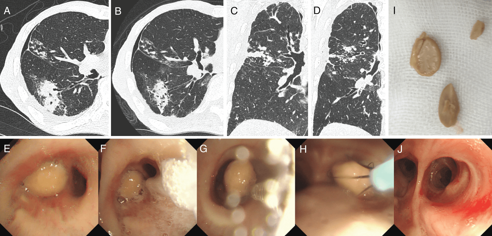
Figure 1. Chest computed tomography (CT) and bronchoscopic findings. (A) Axial and (C) coronal reconstruction CT on admission revealed granular shadows indicative of non-tuberculous mycobacteria in the middle lobe and consolidation attributed to bacterial pneumonia in the right lower lobe. (B) Axial and (D) coronal reconstruction CT demonstrated a new nodular shadow within the right lower lobe bronchus, prompting suspicion of an aspirated boiled soybean. (E) Flexible bronchoscopy revealed an obstructing boiled soybean surrounded by edema and redness. (F) Initially, a brush was used to create a gap, (G) followed by careful extraction using curettage, and (H) basket forceps. (I) The extracted boiled soybean was brittle and fragmented into three pieces. (J) Post-removal examination of the bronchus revealed no residual soybeans.
Discussion
According to reports, foreign body aspiration requiring bronchoscopic intervention is a significant concern, particularly among pediatric patients and older adults, with approximately 40% of cases occurring in adults.3 Commonly encountered foreign bodies include teeth (37.7%), bones (24.6%), and soft organic materials such as boiled soybeans (14.5%),4 presenting a diverse range of challenges for removal.
FB is the preferred method for foreign body removal in adults due to its high success rate, which ranges from 61% to 100%.5 However, the efficacy of this procedure is influenced by various factors, including the patient’s condition, the location and duration of foreign body stagnation, and the foreign body’s physical characteristics, such as size and fragility.3 Dysphagia is a common complication in over 40% of elderly patients with foreign body aspiration,2,4 potentially resulting in a delayed diagnosis. Patient delay in seeking medical attention can lead to progressive granulation and bronchial stenosis, thereby increasing the risk of procedural complications.2,4,6 Although FB was attempted shortly after aspiration of a boiled soybean in the presented case, the soybean adhered to the bronchial mucosa more firmly than anticipated. This condition required careful extraction using multiple devices to prevent bleeding. The high vegetable fat content of the soybean likely induced significant inflammation and mucosal edema. Additionally, water absorption led to swelling, rendering the soybean brittle and prone to fragmentation, thereby complicating its retrieval. The success of foreign body removal depends on the timing of the aspiration for the removal procedure and the inflammatory effects induced by the foreign body.
Curettage was used for foreign body removal, though recent advancements in bronchoscopic devices provide various equipment options. Cryoextraction, a technique that has advanced significantly in recent years, was also considered a viable option. However, concerns arose regarding potential damage to the surrounding bronchial tissue, given the 10 mm lumen in the right basal bronchus. Moreover, the cost burden of this process is substantial, and the number of facilities able to adopt this procedure is limited. We also considered prioritizing other forceps, such as Zero Tip and tripod grasping forceps, but we opted for curettage due to the fragility of boiled soybeans. Indeed, the boiled soybean quickly crumbled during the final retrieval attempt using basket forceps (Fig. 1I and video file). Fragmentation could have occurred if basket forceps were utilized while the soybean adhered to the mucosa, potentially resulting in retrieval failure.
Bronchoscopic balloon catheters are another option, but their effectiveness depends on factors like the balloon size relative to the bronchial lumen, the foreign body’s fragility, and adherence to the bronchial mucosa. Due to the boiled soybean’s 10 mm diameter, similar to the length of the curette from joint to tip, and its 2 mm width, we anticipated minimal mucosal damage with curettage. Curettage also offered the largest contact area with the boiled soybean among the available devices, minimizing the risk of collapse. Given the high cost of balloon catheters as opposed to that of curettage, we opted for the latter as the preferred removal method.
Considering the nature of foreign body removal in this case, it is crucial to carefully assess potential adverse events associated with curettage, including airway mucosal injury, bleeding, fragmentation leading to retrieval failure, and displacement of the foreign body.
Moreover, the limited experience with FB for foreign body removal poses a challenge. Sancho-Chust et al. reported that only 0.25% of all FB procedures involved foreign body removal,6 underscoring the need for enhanced training methods. With limited information on whether curettage is optimal for boiled soybean removal, additional cases are needed for future research. Video demonstrations provide a clearer view of the removal process than static images, offering valuable insights into techniques and foreign body conditions. Sharing simulated experiences via video may significantly enhance procedural proficiency, as video simulations offer greater educational benefits compared to static images.
Conclusions
This case represents an opportunity to simulate the removal of a boiled soybean using FB through video files. Future implementation of virtual bronchoscopy training based on such simulations is expected to enhance the success rate of FB for foreign body removal.
Acknowledgement
We appreciate the patient’s cooperation in the data collection.
Funding Source
No financial support was provided.
Author Contributions
S.M., N.T., R.O., A.M., M.K., K.Y., H.K., and K.T. were responsible for data curation. F.K. and K.K. interpreted the data and drafted the original manuscript. H.K. and K.T. made substantial contributions to revising the manuscript drafts. All authors have read the manuscript and agree with the content and data.
Data Availability Statement
The datasets used in the current study are available from the corresponding author upon reasonable request.
Ethical Statement
The article does not involve the participation of any animals. A written informed consent was obtained from the patient for the publication of this report and accompanying images.
Conflict of Interest
The authors report no conflicts of interest in this work.
Supplemental Information
Supplemental information for this article can be found online at https://sup.jclinque.com/api/articles/37/download-suppl.
References
[1] Ma W, Hu J, Yang M, et al. Application of flexible fiberoptic bronchoscopy in the removal of adult airway foreign bodies. BMC Surg. July 23, 2020;20(1):165. doi: 10.1186/s12893-020-00825-5.
[2] Costa ADS Jr., Palomino ALM, Suzuki I, et al. Analysis of 108 flexible bronchoscopies for the removal of foreign bodies from the airways. Einstein (Sao Paulo). 2023;21(10):eAO0391. doi: 10.31744/einstein_journal/2023AO0391.
[3] Ünal G, Yılmaz A, Tok T, et al. Use of flexible bronchoscopy in foreign body aspiration. Turk Arch Otorhinolaryngol. June 2022;60(2):88–94. doi: 10.4274/tao.2022.2022-6-6.
[4] Jang G, Song JW, Kim HJ, et al. Foreign-body aspiration into the lower airways in adults; multicenter study. PLoS One. 2022;17(7):e0269493. doi: 10.1371/journal.pone.0269493.
[5] Sehgal IS, Dhooria S, Ram B, et al. Foreign body inhalation in the adult population: experience of 25,998 bronchoscopies and systematic review of the literature. Respir Care. October 2015;60(10):1438–1448. doi: 10.4187/respcare.03976.
[6] Sancho-Chust JN, Molina V, Vañes S, et al. Utility of flexible bronchoscopy for airway foreign bodies removal in adults. J Clin Med. May 10, 2020;9(5):1409. doi: 10.3390/jcm9051409.
 |
Copyright: © 2024 Kashizaki et al. This work is licensed under a Creative Commons Attribution 4.0 International License, which permits unrestricted use, distribution, and reproduction in any medium, provided the original work is properly cited. |