| Journal of Clinical Question. 2025; 2(6): e96 https://doi.org/10.69854/jcq.2025.0035 Advance access publication date 03 December 2025 |
 |
Case Report
Remote Cerebellar Hemorrhage following Surgery for Ossification of the Ligamentum Flavum at the Upper Thoracic Spine: A Case Report and Literature Review
1Department of Spinal Surgery, Heiwa Hospital, Yokohama, Japan.
2Department of Orthopedic Surgery, Makino Memorial Hospital, Yokohama, Japan.
*Corresponding Author: e-mail: youngcmu@gmail.com
Submitted: October 02, 2025 Accepted: December 02, 2025
Clinical Question Box
Which clinical factors are associated with the choice of conservative versus interventional management in patients with remote cerebellar hemorrhage?
An analysis of patient characteristics revealed no significant associations between demographic or clinical factors and the chosen management approach. Variables such as age, sex, type of durotomy, hypertension, diabetes mellitus, and anticoagulant use did not differ meaningfully between patients managed conservatively and those who required intervention. The decision to pursue conservative or interventional management in cases of remote cerebellar hemorrhage appears to be guided primarily by clinical presentation and radiological severity rather than by patient demographics or comorbidities.
Abstract
Introduction: Remote cerebellar hemorrhage (RCH) is a rare but serious complication following cranial or spinal surgery, typically attributed to excessive cerebrospinal fluid (CSF) loss and subsequent venous tearing. Although uncommon after spinal procedures, RCH warrants prompt recognition, particularly when dural injury occurs during decompression for ossification of the ligamentum flavum (OLF). Case Presentation: A 73-year-old man with hypertension and diffuse idiopathic skeletal hyperostosis (DISH) underwent T1–T2 decompression for upper thoracic OLF. An intraoperative dural tear with CSF leakage was repaired; however, the patient developed postoperative seizures, and imaging revealed bilateral cerebellar hemorrhages. Conservative management, including drainage adjustment and secondary dural repair, resulted in full neurological recovery and complete radiological resolution by postoperative day 34. Conclusion: RCH should be considered in patients presenting with neurological deterioration following spinal surgery complicated by CSF leakage. The coexistence of OLF and DISH increases surgical complexity and may elevate the risk of RCH. Meticulous dural handling and careful CSF management are essential to prevent this rare but potentially serious complication.
Keywords: Remote cerebellar hemorrhage, ossification of ligamentum flavum, cerebrospinal fluid, clinical feature, case report
Introduction
Remote cerebellar hemorrhage (RCH) is a rare but potentially serious postoperative complication that typically occurs after supratentorial or spinal surgery.1 The first case was reported in 1981 following a cervical laminectomy performed in the sitting position.2 RCH is characterized by hemorrhage within the cerebellum, remote from the surgical site, and is most commonly associated with excessive cerebrospinal fluid (CSF) loss during or after surgery. This loss of CSF can lead to downward displacement of the cerebellum and subsequent tearing of bridging veins.3 Although the incidence of RCH following spinal procedures is exceedingly low, awareness of this condition is essential for timely diagnosis and appropriate management.
Ossification of the ligamentum flavum (OLF) is a pathological condition characterized by ectopic bone formation within the ligamentum flavum, resulting in spinal canal stenosis and myelopathy, particularly in the thoracic region.4 Surgical decompression remains the mainstay of treatment for symptomatic OLF; however, procedures involving the upper thoracic levels carry inherent risks due to anatomical complexity and the proximity of the dura mater.5 Dural tears and CSF leakage are well-recognized intraoperative complications of OLF surgery, and these events have been implicated as potential precipitants of postoperative RCH.6 Here, we report a rare case of RCH occurring after decompressive surgery for upper thoracic OLF.
Case Presentation
A 73-year-old man with hypertension and a history of lacunar infarction presented with bilateral lower extremity numbness, gait instability, and recurrent falls. Brain magnetic resonance imaging (MRI) revealed no intracranial abnormalities; however, cervical and thoracic MRI demonstrated marked OLF at the T1–T2 level, resulting in severe spinal cord compression. Computed tomography (CT) further showed diffuse idiopathic skeletal hyperostosis (DISH) extending from C2–C7 and T2–T12, with relative sparing of the C7–T2 segment (Fig. 1A,1B). After discontinuation of aspirin 5 days before surgery, the patient underwent T1–T2 laminectomy with pedicle screw fixation at C7–T2. During OLF removal, a right-sided dural tear occurred, leading to CSF leakage, which was repaired using a Neoveil® sheet and Bolheal®. An SB-VAC® negative-pressure drain was placed in the extradural space.
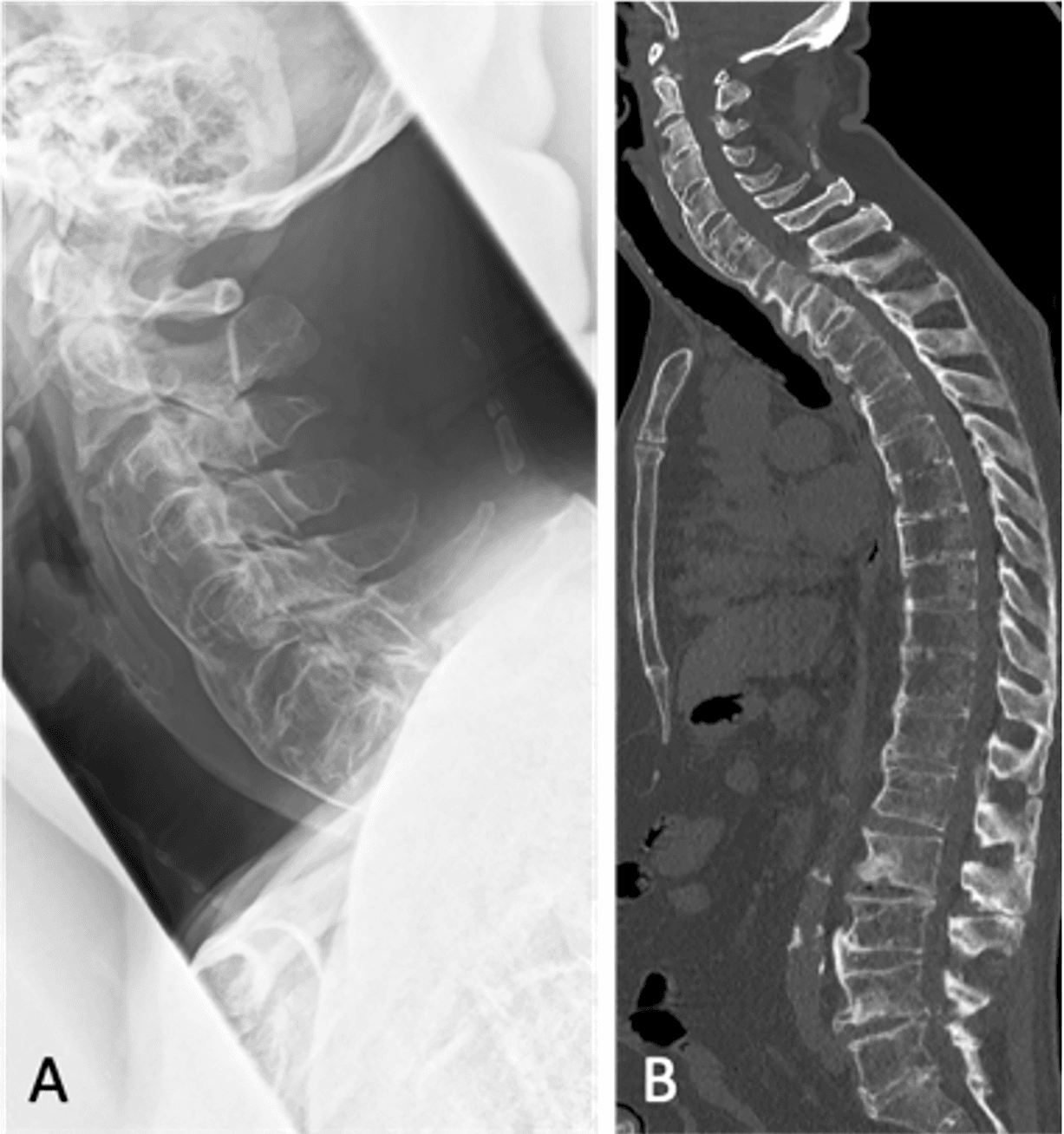
Figure 1. Preoperative imaging of OLF and diffuse idiopathic skeletal hyperostosis. (A) Lateral cervical spine X-ray and (B) whole-spine CT scan. CT, computed tomography; OLF, ossification of the ligamentum flavum.
Two hours postoperatively, 160 mL of drainage was recorded without evidence of CSF leakage, and the drainage pressure was reduced to half. Approximately 6 hours later, the patient developed a generalized tonic seizure during repositioning, accompanied by a sudden increase in drainage output that was consistent with CSF in appearance. Brain CT revealed a right cerebellar hemorrhage (CBH) and subarachnoid hemorrhage (Fig. 2). The Glasgow Coma Scale score was 8 (E2V2M4) without motor weakness. Management included converting the drain to natural pressure 12 hours postoperatively. On postoperative day 1 (POD1), brain CT demonstrated an increase in the size of the right CBH (Fig. 3A). Aspirin was not reinitiated due to the substantial risk of intracranial hemorrhage. By POD 3, follow-up CT showed bilateral CBH (Fig. 3B), prompting drain removal. Cerebral angiography performed on POD 5 revealed no vascular abnormalities. MRI confirmed persistent CSF leakage and subcutaneous fluid collection (Fig. 4A, 4B). Consequently, dural repair and wound closure were performed under local anesthesia with intravenous sedation on POD 7.
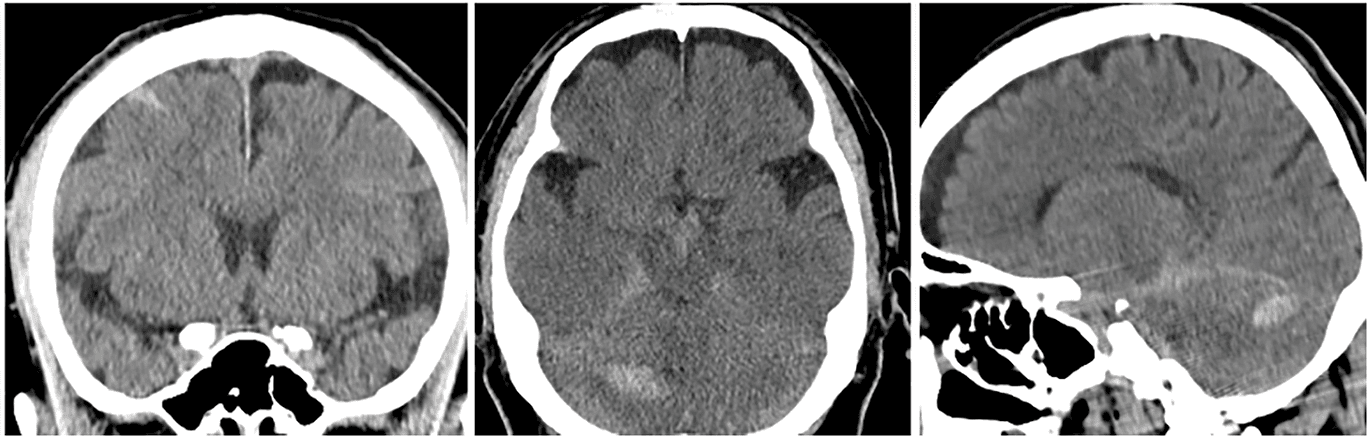
Figure 2. Postoperative cerebellar hemorrhage. Brain CT demonstrating a right cerebellar linear hemorrhage and subarachnoid hemorrhage following acute CSF loss. CSF, cerebrospinal fluid; CT, computed tomography.
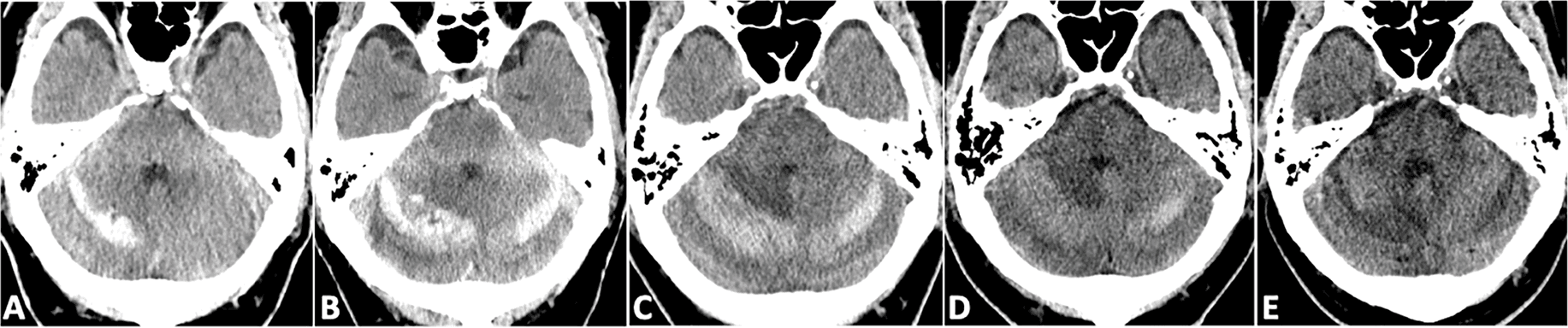
Figure 3. Serial brain CT changes of cerebellar hemorrhage. (A) POD 1: Increased right cerebellar hemorrhage; (B) POD 3: Bilateral cerebellar hemorrhages; (C) POD 7: No further hemorrhagic progression; (D) POD 10: Partial resolution of hemorrhages; (E) POD 34: Complete resolution of cerebellar hemorrhages. CT, computed tomography; POD, postoperative day.
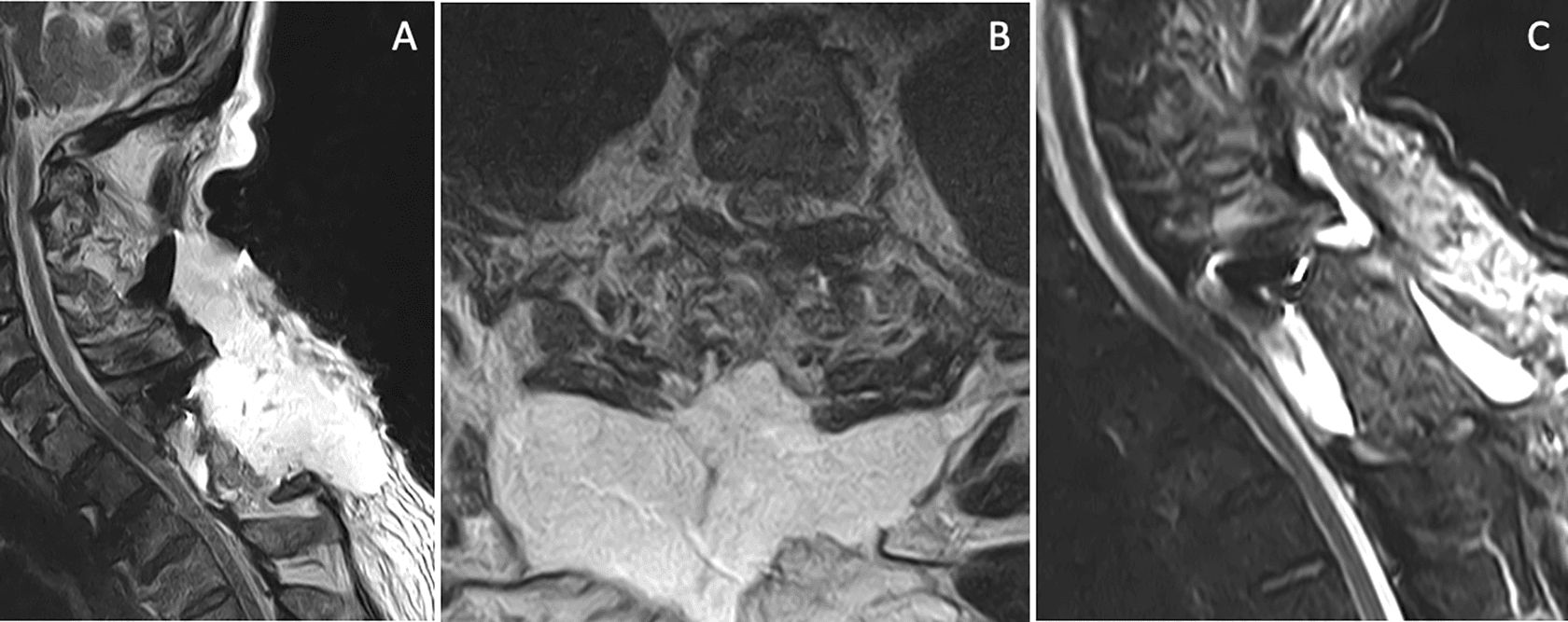
Figure 4. CSF leakage and post-repair outcome. (A, B) MRI showing subcutaneous CSF accumulation and persistent leakage before repair; (C) POD 34 MRI confirming complete resolution of CSF leakage. CSF, cerebrospinal fluid; MRI, magnetic resonance imaging; POD, postoperative day.
Following the procedure, the patient’s level of consciousness improved markedly. On POD 10, CT imaging demonstrated partial resolution of the CBH (Fig. 3D), and the patient regained ambulatory function with the assistance of a walker. After 2 weeks of rehabilitation, he achieved a stable gait and remained free of headaches. By POD 34, imaging confirmed complete resolution of the hemorrhages (Fig. 3E) and the absence of CSF leakage (Fig. 4C). The patient was discharged 8 weeks postoperatively, ambulatory and without evidence of ataxia. The full course of management is presented in a detailed timeline (Table S1). During the 18-month postoperative follow-up, the patient remained asymptomatic with respect to both the OLF and the RCH complications.
Discussion
The reported incidence of RCH ranges from 0.08% to 0.6%, although the true frequency is likely higher due to underrecognition and underreporting.7–9 RCH may occur immediately after surgery or within several postoperative days. Common clinical manifestations include headache, altered consciousness, nausea, vomiting, dysarthria, ataxia, and motor deficits; seizures are uncommon but characteristic when present.10 The proposed pathophysiological mechanism involves cortical exposure to hemoglobin from subdural bleeding, which exerts proconvulsive effects and typically produces generalized rather than focal seizures. The principal risk factor is dural tear or CSF leakage, often resulting in bilateral cerebellar hemorrhages in the absence of vascular abnormalities. A rapid reduction in intracranial pressure creates a craniospinal pressure gradient that induces cerebellar descent, venous traction, and subsequent hemorrhage. The use of negative-pressure drainage systems further increases this risk. Even with watertight dural closure confirmed by the Valsalva maneuver, microleakage may persist along suture lines.11 Additional factors, such as hypertension and prior cerebrovascular disease, may contribute to vascular fragility. Adjusting the patient’s position (e.g., Trendelenburg) or elevating the drainage reservoir can safely reduce CSF outflow12
To achieve a comprehensive understanding of the characteristics of RCH, we conducted a systematic PubMed search on October 1, 2025, using the keywords “remote cerebellar hemorrhage” and “spinal surgery,” along with an updated review based on the work of Allouch et al.13 All data were extracted and screened by two authors. The clinical characteristics of 90 patients with RCH are summarized in Table 1.13–20 The mean age of patients was 60.2 ± 16.1 years, with a slight female predominance (53/90, 59%).
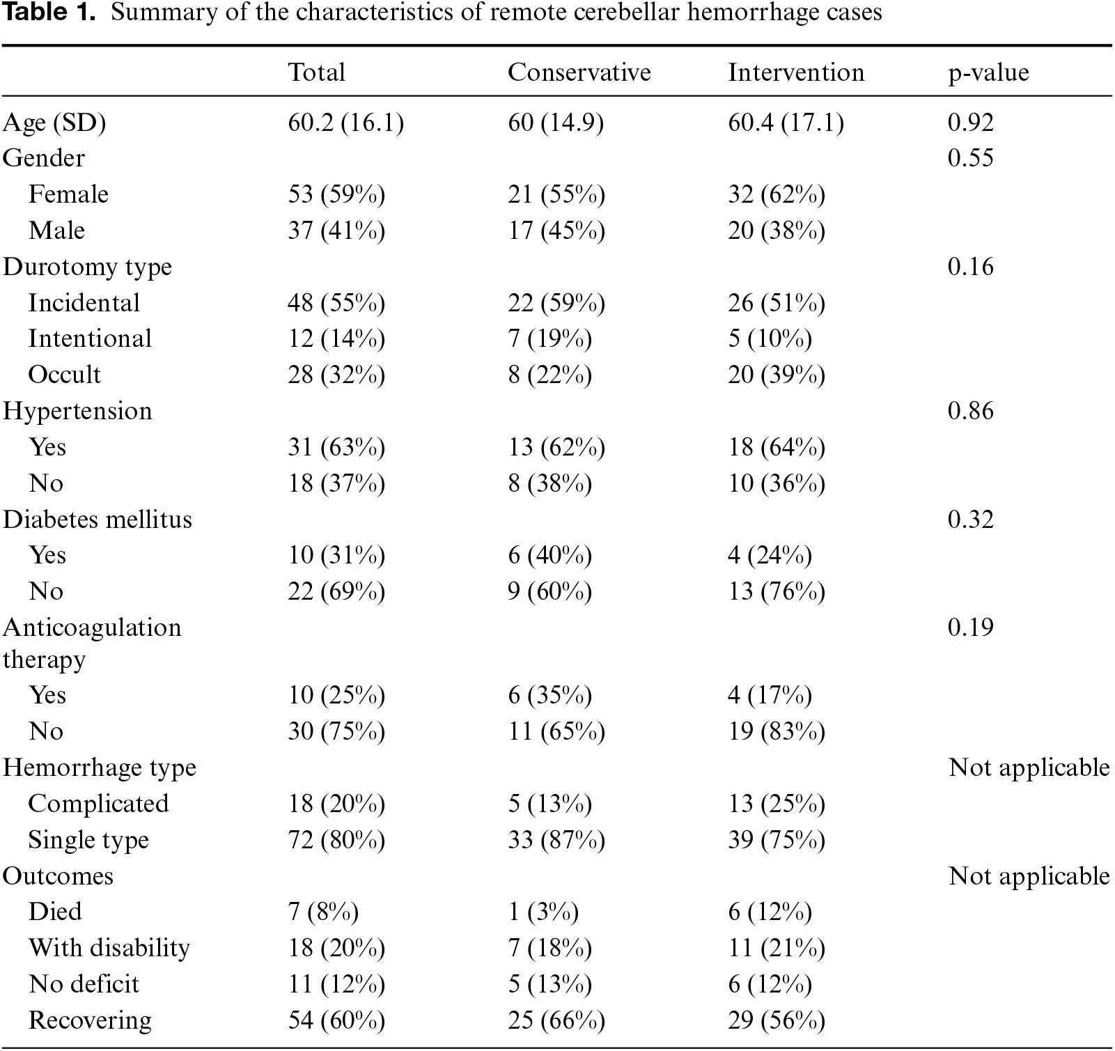
Among the cases, incidental durotomy was the most common type (48/90, 55%), followed by occult durotomy (28/90, 31.8%) and intentional durotomy (12/90, 13.6%). A single type of hemorrhage was observed in 65 of 90 cases (80%), with CBH accounting for 43 of 72 cases (59.7%). Complicated hemorrhages occurred in 20% of cases, all of which were associated with CBH. Comorbid hypertension was present in 31 of 49 patients (63%), and diabetes mellitus in 10 of 32 patients (31%). Regarding anticoagulation status, 10 of 40 patients (25%) were receiving anticoagulant therapy. Management was predominantly conservative in 38 of 90 cases (42%), while 34 of 90 cases (38%) required a single intervention, and 18 of 90 cases (20%) required multiple interventions. In terms of outcomes, 7 of 90 patients (8%) died, 18 of 90 (20%) had residual disability, and the remaining patients achieved good recovery. Age, sex, durotomy type, and comorbidities showed no statistically significant association with the choice between conservative and interventional management for RCH.
OLF predominantly affects the lower thoracic spine in approximately 67% of cases, whereas only about 6% occur in the upper thoracic region. DISH is observed in nearly 21% of OLF cases, most commonly involving the thoracolumbar spine.21 Upper thoracic OLF associated with DISH is exceedingly rare, with only two cases reported to date—both presenting with concurrent ossification of the posterior longitudinal ligament.22,23 In the present case, the coexistence of OLF and DISH increases the technical complexity of spinal surgery and heightens the risk of dural tears with CSF leakage, both of which are recognized predisposing factors for RCH. Additional risk factors include advanced age, hypertension, antiplatelet therapy, and excessive intraoperative CSF drainage or negative-pressure suction. These anatomical and physiological considerations underscore the importance of meticulous dural handling, judicious CSF management, and vigilant postoperative monitoring to minimize the risk of RCH, particularly in patients with OLF associated with DISH.
Conclusion
RCH is a rare but serious complication following spinal surgery, typically associated with CSF loss due to dural tears. This case highlights the potential risk of RCH after decompression for upper thoracic ossification of the OLF in the presence of DISH. The coexistence of OLF and DISH increases surgical complexity and the likelihood of CSF leakage. Meticulous dural management, controlled drainage, and vigilant postoperative monitoring are crucial to preventing RCH and ensuring favorable patient outcomes.
Acknowledgment
We appreciate the patient’s cooperation in data collection.
Funding Source
No financial support was provided.
Author Contributions
B.Y. was responsible for data curation, data interpretation, and drafting of the original manuscript. T.S., M.T., O.K., and Y.O. were responsible for the revision. All authors have read and approved the manuscript and agree with the content and data.
Data Availability Statement
The datasets used in the current study are available from the corresponding author upon reasonable request.
Ethical Statement
This article does not involve the participation of any animals. The patient gave written informed consent for the publication of this report and accompanying images.
Conflict of Interest
The authors report no conflicts of interest in this work.
Supplemental Information
Supplemental information for this article can be found online at https://sup.jclinque.com/api/articles/96/download-suppl.
References
[1] Narita H, Narikiyo M, Hirokawa Y, et al. Case of fatal cerebellar hemorrhage following lumbar spinal surgery with dural leak. Surg Neurol Int. 2025;16:312. doi:10.25259/sni_608_2025.
[2] Chadduck WM. Cerebellar hemorrhage complicating cervical laminectomy. Neurosurgery. August 1981;9(2):185–189. doi:10.1227/00006123-198108000-00016.
[3] Di L, Wei G, Eichberg DG, Komotar RJ, Ivan M. Remote cerebellar hemorrhage associated with intra-operative cerebrospinal fluid leak: a report of two rare case presentations and review of the literature. Cureus. December 14, 2020;12(12):e12082. doi:10.7759/cureus.12082.
[4] Devani K, Purandare A, Wankhade R, Palave P, Sharma A, Diyora B. Coexisting spinal enthesopathy syndromes–A rare finding. J Spinal Surg. 2022;9(3):173–177. doi:10.4103/joss.joss_10_22.
[5] Mensah EO, Chalif JI, Baker JG, Chalif E, Biundo J, Groff MW. Challenges in contemporary spine surgery: a comprehensive review of surgical, technological, and patient-specific issues. J Clin Med. September 14, 2024;13(18):5460. doi:10.3390/jcm13185460.
[6] Milton R, Kalanjiyam GP, Rajasekaran S, Shetty AP, Kanna RM. Dural injury following elective spine surgery-A prospective analysis of risk factors, management and complications. J Clin Orthop Trauma. June 2023;41(3):102172. doi:10.1016/j.jcot.2023.102172.
[7] Yan X, Yan LR, Ma ZG, et al. Clinical characteristics and risk factors of intracranial hemorrhage after spinal surgery. World J Clin Cases. August 16, 2023;11(23):5430–5439. doi:10.12998/wjcc.v11.i23.5430.
[8] Kaloostian PE, Kim JE, Bydon A, et al. Intracranial hemorrhage after spine surgery. J Neurosurg Spine. September 2013;19(3):370–380. doi:10.3171/2013.6.Spine12863.
[9] Gerstmeyer J, Avantaggio A, Pierre C, et al. The hidden risk: intracranial hemorrhage following durotomies in spine surgery. North American Spine Soc J (NASSJ). 2024;20(2):100555. doi:10.1016/j.xnsj.2024.100555.
[10] Bashti M, DeLong C, Shah S, Di L, Jaman E, Basil GW. Postoperative remote cerebellar hemorrhage following lumbar laminectomy without cerebrospinal fluid leak: a rare presentation and review of the literature. Interdiscip Neurosurg. June 01, 2024;36:101908. doi:10.1016/j.inat.2023.101908.
[11] Chan AY, Mullin JP, Benzel E, Bingaman W. Immediate remote cerebellar hemorrhage and seizure following revision lumbosacral fusion. Cureus. May 30, 2017;9(5):e1292. doi:10.7759/cureus.1292.
[12] Dong P, Huang J, Deng X, Yang H, Luo C. The drainage volume control by elevation of drainage height versus head down tilt in supine position for management of cerebrospinal fluid leakage following lumbar posterior surgery. BMC Musculoskelet Disord. November 14, 2024;25(1):910. doi:10.1186/s12891-024-08040-6.
[13] Allouch H, Abu Nahleh K, Mursch K, Shousha M, Alhashash M, Boehm H. Symptomatic intracranial hemorrhage after dural tear in spinal surgery-a series of 10 cases and review of the literature. World Neurosurg. June 2021;150:e52–e65. doi:10.1016/j.wneu.2021.02.071.
[14] Hofler RC, Wemhoff MP, Johans SJ, Nockels RP. Cerebellar hemorrhage following an uncomplicated lumbar spine surgery: case report. J Stroke Cerebrovasc Dis. July 2019;28(7):e104–e105. doi:10.1016/j.jstrokecerebrovasdis.2018.12.001.
[15] Kinthala S, Jiao K, Ankam A, Paramore CG. Cerebellar hemorrhage and spinal fluid overdrainage with tonsillar herniation following spine surgery. Cureus. September 13, 2020;12(9):e10418. doi:10.7759/cureus.10418.
[16] Lim CHS, Salkade PR, Peter AC. Remote cerebellar hemorrhage as a complication of lumbar spine surgery. J Radiol Case Rep. February 2020;14(2):1–11. doi:10.3941/jrcr.v14i2.3844.
[17] Sánchez Zamora P, Gómez Del Pulgar Vázquez B, Gholamian Ovejero S. Intraventricular hemorrhage as complication after spinal surgery. Case report. Rev Esp Anestesiol Reanim (Engl Ed). March 2022;69(3):183–186. doi:10.1016/j.redare.2022.02.003.
[18] Sato S, Hayashi H, Urayama D, Ito Y, Naganuma H, Shinbori H. A case of remote cerebellar hemorrhage in spinal surgery without dural injury: case report. J Orthop Case Rep. September 2023;13(9):38–41. doi:10.13107/jocr.2023.v13.i09.3868.
[19] Pappa E, Pilichou A, Antonopoulos SI, et al. Surgical treatment of congenital kyphosis in children: report of a rare complication of remote cerebellar haemorrhage. Cureus. March 2024;16(3):e56488. doi:10.7759/cureus.56488.
[20] Obeng-Gyasi B, Love ES, Swar-Eldahab M, Blackwell M, Chinthala AS, Mao G. Hidden hazard of remote cerebellar hemorrhage after cervical spine surgery: case report. Int J Surg Case Rep. August 2025;133:111571. doi:10.1016/j.ijscr.2025.111571.
[21] Ehara S, Shimamura T, Nakamura R, Yamazaki K. Paravertebral ligamentous ossification: DISH, OPLL and OLF. Eur J Radiol. July 1998;27(3):196–205. doi:10.1016/s0720-048x(97)00164-2.
[22] Alijani B, Yousefzadeh-Chabok S, Ramzannejad A, Behzadnia H, Emamhadi M. Simultaneous diffuse idiopathic hyperostosis, ossification of the posterior longitudinal ligament and ligamentum flavum. Case report. Iranian J Neurosurg. 2018;4(1):51–55. doi:10.32598/irjns.4.1.51.
[23] Guo Q, Ni B, Yang J, Zhu Z, Yang J. Simultaneous ossification of the posterior longitudinal ligament and ossification of the ligamentum flavum causing upper thoracic myelopathy in DISH: case report and literature review. Eur Spine J. July 2011;20 Suppl 2(Suppl 2):S195–S201. doi:10.1007/s00586-010-1538-z.
 |
Copyright: © 2025 Yang et al. This work is licensed under a Creative Commons Attribution 4.0 International License, which permits unrestricted use, distribution, and reproduction in any medium, provided the original work is properly cited. |