| Journal of Clinical Question. 2026; 3(4): e369 https://doi.org/10.69854/jcq.2026.0021 Advance access publication date 18 August 2026 |
 |
Review
Factors Influencing the Efficacy of Programmed Cell Death Protein 1/Programmed Death-Ligand 1 Inhibitors in Non-Small Cell Lung Cancer
Long-Hua Lu1, Ze-Yang Zeng1, Zi-Qi Guan1, Luan Wang2,*, Xiao-Ying Qi3,*, Shu-Ling Zhang 3,*
3,*
1Department of Clinical Medicine, China Medical University, Shenyang, 110122, China.
2Department of Oncology, Shengjing Hospital of China Medical University, Shenyang 110004, China.
3Department of Emergency Medicine, Shengjing Hospital of China Medical University, Shenyang 110004, China.
*Corresponding Authors: L.W.: wangluan@sj-hospital.org; X.Q.: qixiaoying@sj-hospital.org; S.Z.: slzhang@cmu.edu.cn
Submitted: April 25, 2026 Accepted: August 17, 2026
Clinical Question Box
What factors affect the efficacy of immune checkpoint inhibitors (ICIs), especially PD-(L)1 inhibitors, in patients with non-small cell lung cancer?
The efficacy of PD-(L)1 inhibitors in non-small cell lung cancer reflects interactions among tumor biology, the tumor-microenvironment, treatment context, and host factors. Higher tumor-cell PD-L1 expression and an inflamed immune contexture are the most clinically established favorable features. Tumor mutational burden, T-cell states, tertiary lymphoid structures, oncogenic drivers and co-mutations, body composition, microbiota, and concomitant medications may provide additional information, but most remain context-dependent or investigational. Actionable oncogenic drivers, poor performance status, cachexia, and baseline corticosteroid use for cancer-related symptoms are generally associated with reduced benefit, particularly from ICI monotherapy. No single variable is sufficiently accurate for universal treatment selection; integrated and dynamic assessment is therefore required.
Abstract
Immune checkpoint inhibitors (ICIs), particularly programmed cell death protein 1/programmed death-ligand 1 (PD-1/PD-L1) inhibitors, are central to the treatment of non-small cell lung cancer (NSCLC), and a subset of patients achieves durable benefit. In historical studies of broadly selected patients receiving ICI monotherapy, objective responses occurred in approximately 20%, although response rates vary substantially according to treatment line, PD-L1 expression, molecular subtype, patient selection, and the use of combination regimens. This narrative review used targeted searches of PubMed/MEDLINE, ClinicalTrials.gov, and reference lists of key publications to identify English-language evidence available through May 2026 and prioritized pivotal randomized trials, prospective translational studies, consensus statements, guidelines, and recent high-quality reviews. We critically examine tumor-microenvironmental features, PD-L1 expression, tumor mutational burden (TMB), oncogenic driver alterations, combination strategies, and patient-related factors that may influence PD-(L)1 inhibitor efficacy. Although other immune checkpoints, including cytotoxic T-lymphocyte-associated protein 4 and lymphocyte-activation gene 3, are discussed when directly relevant to combination therapy, the principal focus is PD-1/PD-L1-directed treatment. Current evidence supports PD-L1 as the most widely implemented biomarker, but no single factor adequately captures the biological and temporal heterogeneity of treatment response. Integrated, dynamic, and context-specific biomarker models, supported by prospective validation, are therefore required to improve precision immuno-oncology.
Keywords: Non-small cell lung cancer, programmed cell death protein 1, immune checkpoint inhibitors, tumor microenvironment, tumor mutational burden, combination therapy
Background
The programmed cell death protein 1 (PD-1)/programmed death-ligand 1 (PD-L1) axis is a major mechanism of tumor immune escape that attenuates antitumor T-cell activity. ICIs targeting PD-1, including nivolumab and pembrolizumab, and PD-L1, including atezolizumab and durvalumab, have improved survival across multiple stages and treatment settings of NSCLC.1
Immune checkpoint inhibition also encompasses targets such as cytotoxic T-lymphocyte-associated protein 4 (CTLA-4) and lymphocyte-activation gene 3 (LAG-3). However, PD-(L)1 blockade remains the principal immunotherapeutic backbone in NSCLC, whereas CTLA-4, LAG-3, and other checkpoint-directed agents are generally used or investigated in combination with PD-(L)1 inhibitors. Accordingly, this review focuses on factors that influence the efficacy of PD-1/PD-L1-directed therapy; broader immunotherapy concepts are included only when they directly inform this scope.2
The magnitude of benefit from PD-(L)1 blockade is highly context-dependent. Historical ICI monotherapy studies in broadly selected advanced NSCLC populations reported objective response rates of approximately 20%, whereas substantially higher rates are observed in selected patients with high PD-L1 expression and with chemoimmunotherapy or other combination regimens.3 Response also varies by treatment line, histologic and molecular subtype, disease burden, and host condition. Primary and acquired resistance therefore remain common and create an urgent need for clinically reproducible predictive strategies. This review critically evaluates tumor-microenvironmental factors, PD-L1, tumor mutational burden (TMB), oncogenic driver alterations, treatment combinations, and patient characteristics, with particular attention to evidentiary limitations and clinical actionability.
Relevant English-language publications were identified through targeted searches of PubMed/MEDLINE and ClinicalTrials.gov and through review of the reference lists of pivotal articles, with coverage through May 2026. Priority was given to randomized trials, prospective clinical and translational studies, major retrospective cohorts, consensus statements, guidelines, and recent high-quality reviews. Because the objective was an interpretive narrative synthesis rather than an exhaustive systematic review, no PRISMA-based study-selection process or formal risk-of-bias assessment was performed.
Tumor-Microenvironment (TME) Factors
The TME comprises tumor and immune cells, vascular and stromal compartments, signaling molecules, and the extracellular matrix. A commonly used spatial framework classifies tumors as immune-inflamed, immune-excluded, or immune-desert. Immune-inflamed tumors contain intratumoral T cells and interferon-related signaling and are generally more responsive to PD-(L)1 blockade.4 In immune-excluded tumors, CD8+ T cells accumulate at the invasive margin or within stroma without effective tumor penetration, whereas immune-desert tumors contain few tumor-reactive T cells. These phenotypes are biologically useful but are neither discrete nor stable, and their assessment is not standardized; individual tumors may contain mixed regions and may change over time or during treatment.5–7
Clinical response reflects the balance between antitumor immunity and suppressive cellular and stromal programs. CD8+ T-cell recruitment and activation can favor response, whereas MDSCs, Tregs, suppressive myeloid states, N2-like TANs, fibroblast barriers, and abnormal vasculature may limit immune-cell trafficking or function (Fig. 1). Many of these relationships are supported by translational or observational evidence and have not yet yielded standardized tests for routine treatment selection.
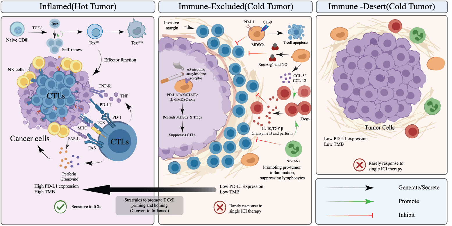
Figure 1. Conceptual framework of immune-inflamed, immune-excluded, and immune-desert tumor microenvironments. Immune-inflamed tumors contain intratumoral effector lymphocytes and are more likely to respond to immune checkpoint blockade, whereas stromal exclusion and paucity of tumor-reactive T cells may limit response. The categories are dynamic and overlapping rather than fixed. Abbreviations: Arg, arginase; α5-nAChR, α5 nicotinic acetylcholine receptor; CCL, C–C motif chemokine ligand; CTL, cytotoxic T lymphocyte; Fas, Fas cell surface death receptor; FasL, Fas ligand; Gal, galectin; ICI, immune checkpoint inhibitor; IL, interleukin; MDSC, myeloid-derived suppressor cell; MHC, major histocompatibility complex; NK, natural killer; NO, nitric oxide; PD-1, programmed cell death protein 1; PD-L1, programmed death-ligand 1; ROS, reactive oxygen species; STAT, signal transducer and activator of transcription; TAM, tumor-associated macrophage; TAN, tumor-associated neutrophil; TCF1, T-cell factor 1; TCR, T-cell receptor; Texeff, effector-exhausted T cell; Texterm, terminally exhausted T cell; TGF, transforming growth factor; TMB, tumor mutational burden; TME, tumor microenvironment; TNF, tumor necrosis factor; Tpex, progenitor-exhausted T cell; Treg, regulatory T cell.
CD8+ T-Cell Infiltration and Immune Activation
CD8+ T cells, or cytotoxic T lymphocytes, eliminate tumor cells after antigen recognition, and greater intratumoral CD8+ T-cell density has been associated with improved outcomes after ICI therapy in NSCLC.8 Functional state is at least as important as cell number. CD28 co-expression may identify a population with preserved proliferative capacity, whereas high expression of inhibitory receptors such as TIM-3 can mark dysfunctional or terminally exhausted states and potential resistance mechanisms.9
Progenitor-exhausted T (Tpex) cells, commonly characterized by TCF1 and PD-1 expression, retain stem-like properties and can self-renew and generate more differentiated effector-exhausted cells after PD-1 blockade.10,11 Higher Tpex-related signatures or TCF1+PD-1+ tumor-infiltrating lymphocytes have been associated with response and survival in NSCLC. However, Tpex identification is not standardized. It may require multiparameter immunohistochemistry, flow cytometry, or high-dimensional single-cell assays; marker definitions, spatial compartments, sampling time, and analytic thresholds differ across studies. Tpex measurement therefore remains investigational and is not currently a routine clinical biomarker.12
Tertiary Lymphoid Structures and High-Resolution TME Profiling
Tertiary lymphoid structures (TLSs) are organized aggregates of B cells, T cells, dendritic cells, and specialized stromal or vascular elements that can support local antigen presentation and lymphocyte activation. Mature, germinal-center-containing TLSs and greater TLS abundance have been associated with pathological response and improved outcomes after immunotherapy or chemoimmunotherapy in NSCLC.13,14 Nevertheless, TLS presence alone is not uniformly predictive; maturation state, location, stromal context, specimen size, and scoring method can alter interpretation.
Single-cell RNA sequencing and spatial transcriptomic or multiplex-imaging approaches can resolve immune-cell states, clonotypes, spatial neighborhoods, and cell–cell communication networks that are obscured by bulk assays. These methods have identified potentially informative interactions among Tpex cells, dendritic cells, B-cell-rich TLSs, macrophages, fibroblasts, endothelial cells, and tumor cells.10,15 Their clinical translation remains limited by tissue requirements, cost, technical variability, computational dependence, and the absence of prospectively validated decision thresholds.
Despite strong biological rationale, most TME biomarkers remain investigational because reproducibility is affected by intratumoral and temporal heterogeneity, biopsy site and size, pre-analytical handling, assay platform, cell-state definitions, spatial scoring, and treatment exposure. Prospective studies using analytically validated assays and predefined thresholds are needed before these measurements can reliably guide therapy.
Immunosuppressive Network
Myeloid-derived suppressor cells (MDSCs): MDSCs comprise heterogeneous populations of immature myeloid cells that suppress T-cell activity through checkpoint-ligand expression, oxidative and nitrosative stress, amino-acid depletion, and immunosuppressive cytokines. Elevated circulating or intratumoral MDSC levels have been associated with inferior outcomes and primary resistance to ICIs in NSCLC, but they are not validated treatment-selection biomarkers.16,17
Under hypoxic or STAT3-active conditions, monocytic MDSCs may differentiate toward tumor-associated macrophage states. Preclinical depletion or reprogramming of MDSCs can enhance checkpoint blockade, but clinical feasibility remains uncertain. Tumor-intrinsic PD-L1 signaling through the JAK/STAT3/IL-6 axis has also been linked to MDSC-mediated immunosuppression in PD-L1-high NSCLC, providing a mechanistic rationale rather than a validated clinical strategy.18–20
Regulatory T cells (Tregs): Tregs suppress the activities of CD8+ T cells and natural killer cells by secreting inhibitory cytokines (e.g., IL-10, TGF-β), granzyme B, and perforin. Spatial analysis revealed that a shortened mean nearest-neighbor distance between Tregs and CD4+ T cells/tumor cells is predictive of a poor prognosis in NSCLC, whereas proximity to CD8+ T cells may indicate local immune activation.15 PD-L1-positive lung cancer stem-like cells may also contribute to an immunosuppressive lymph-node microenvironment.21
Smoking-related nicotinic receptor signaling may stabilize tumor-cell PD-L1 through STAT3/Jab1 signaling; however, the cited evidence does not establish a direct clinical relationship among smoking, Treg function, and ICI efficacy.22 Experimental strategies that reduce Treg-mediated suppression, including epigenetic modulation, can enhance antitumor immunity in preclinical models, but remain investigational.23
Tumor-associated neutrophils and systemic inflammatory indices: The N1/N2 framework is a useful simplification, but neutrophil states are plastic and cannot be reliably assigned by routine clinical assays. An elevated baseline or early on-treatment neutrophil-to-lymphocyte ratio and platelet-related inflammatory indices are consistently associated with poorer outcomes during ICI-based therapy.24–26 These markers are inexpensive and accessible, but cutoff values vary, and confounding by infection, corticosteroid exposure, tumor burden, and comorbidity limits their use as treatment-specific predictive biomarkers. The reported association between a higher neutrophil-to-lymphocyte ratio and ICI-related myocarditis requires external validation and should not be used for clinical risk stratification at present.27
Cancer Cell-Related Factors
PD-L1 Expression
PD-L1 immunohistochemistry remains the most widely implemented biomarker for selecting PD-(L)1-directed therapy in NSCLC, despite substantial limitations.1,28 Higher tumor-cell PD-L1 expression enriches for response to pembrolizumab monotherapy in oncogene driver-negative advanced NSCLC, but PD-L1 is neither necessary nor sufficient for benefit, particularly when ICIs are combined with chemotherapy.
PD-L1 is spatially and temporally heterogeneous and can be altered by prior therapy, inflammation, specimen type, and sampling site. Patients with low or undetectable expression may respond, whereas some tumors with high expression exhibit primary resistance.28,29 Small biopsies and cytology specimens may not represent the entire tumor, and interobserver variation near clinical cutoffs can change treatment classification.
Assay selection and scoring method also affect interpretation. The 22C3, 28-8, and SP263 assays generally show comparable tumor-cell staining, whereas SP142 tends to label fewer tumor cells and may classify some specimens at a lower expression category.30 The Tumor Proportion Score (TPS) is the percentage of viable tumor cells with membranous PD-L1 staining and is the principal scoring system used for most NSCLC treatment decisions. The Combined Positive Score (CPS) includes PD-L1-positive tumor cells, lymphocytes, and macrophages relative to the number of viable tumor cells; it is established in several other tumor types but is not the dominant scoring system for NSCLC. Assays, antibodies, platforms, specimen requirements, and clinically validated cutoffs should therefore be matched to the treatment indication rather than assumed to be universally interchangeable. The cellular source of PD-L1 may add biological information, but immune-cell and spatial scoring remain less standardized than tumor-cell TPS.31
Tumor Mutational Burden (TMB)
TMB is generally defined as the number of somatic mutations detected per megabase of interrogated genomic sequence. Higher TMB may increase the probability of generating immunogenic neoantigens, but only a subset of nonsynonymous mutations is processed, presented by major histocompatibility complex molecules, and recognized by T cells. TMB should therefore be regarded as an indirect measure of potential tumor immunogenicity rather than a direct measure of neoantigen quality.32
Feasibility of Predicting Efficacy in NSCLC
A higher TMB can increase the probability of neoantigen generation and has been associated with response to PD-(L)1 inhibitors in several NSCLC cohorts.33–36 However, the randomized evidence is not uniformly positive. CheckMate 026 did not meet its primary progression-free survival endpoint in the prespecified PD-L1-selected population; an exploratory whole-exome analysis suggested improved response and progression-free survival with nivolumab in tumors with high TMB, but not a clear overall survival advantage.37 CheckMate 227 subsequently demonstrated a progression-free survival benefit from nivolumab plus ipilimumab in a TMB-high subgroup, whereas longer-term overall survival benefit was not restricted to that subgroup.38 These findings reduced enthusiasm for using TMB as an isolated treatment-selection biomarker in NSCLC.
TMB is influenced by smoking-related mutagenesis, DNA-repair defects, tumor purity, sequencing depth, and specific genomic contexts. dMMR/MSI-H tumors can accumulate mutations and respond to checkpoint blockade, but MSI-H is uncommon in NSCLC and does not account for most TMB-high cases.38–40 A high TMB may therefore reflect several biologically distinct processes, and its predictive value varies according to tumor type, treatment regimen, and the accompanying immune context.
Challenges in TMB Standardization
Whole-exome sequencing is a research reference method for TMB quantification but is costly, data-intensive, and difficult to implement routinely. Clinical testing generally relies on targeted next-generation sequencing panels. Panel size, genomic territory, tumor purity, germline filtering, inclusion of synonymous variants, driver-variant handling, and bioinformatic pipelines can each shift the reported TMB value. Consequently, the same numerical cutoff cannot be assumed to have identical meaning across assays.
FDA-authorized assays such as FoundationOne CDx and MSK-IMPACT cover different genes and genomic territories and apply different analytic procedures. A designation of 10 mutations/Mb on one panel is therefore not necessarily equivalent to 10 mutations/Mb on another. Pre-analytical variables further affect tissue TMB. Blood TMB is attractive because it is minimally invasive, but low circulating tumor DNA shedding, clonal hematopoiesis, disease burden, and tumor-site heterogeneity can impair accuracy, and tissue-blood concordance has not been consistently validated for treatment selection in phase III NSCLC trials.41
Harmonization initiatives have improved cross-platform understanding, but clinically important disagreement persists near cutoff values and can lead to patient misclassification.42,43 Regulatory and clinical adoption has therefore remained limited relative to early expectations. Negative or equivocal randomized findings, assay dependence, uncertain optimal thresholds, limited reproducibility of blood-based measurements, and incomplete evidence that TMB adds independent information beyond PD-L1 and immune contexture all contribute to this caution.
Future use of TMB should emphasize assay-specific validation, cancer- and treatment-context-specific calibration, and analysis as a continuous or probabilistic variable rather than a universal binary threshold. TMB is most plausibly informative as one component of a composite model incorporating PD-L1, immune-cell or gene-expression signatures, spatial immune architecture, relevant driver and co-mutations, and dynamic blood-based markers.44,45 Such models require prospective validation and demonstration that their use improves clinical outcomes.
Accordingly, future standards should define assay-, tumor-, and treatment-specific interpretation and specify the clinical settings in which tissue- or blood-based TMB measurements are appropriate (Fig. 2).
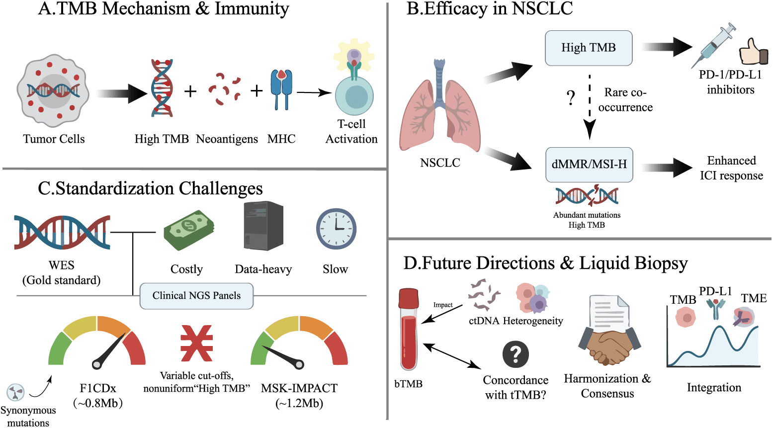
Figure 2. Tumor mutational burden (TMB) as a candidate biomarker in NSCLC. (A) Somatic nonsynonymous mutations can generate neoantigens that are presented through MHC molecules and recognized by T cells. (B) Higher TMB may enrich for response to ICIs, but its predictive value is context dependent; dMMR/MSI-H is uncommon in NSCLC. (C) Whole-exome sequencing is a research reference method, whereas clinical next-generation sequencing panels differ in genomic coverage and bioinformatic methods. (D) Tissue and blood TMB require assay-specific calibration, harmonization, and prospective validation. Abbreviations: bTMB, blood-based tumor mutational burden; ctDNA, circulating tumor DNA; dMMR, deficient mismatch repair; ICI, immune checkpoint inhibitor; MHC, major histocompatibility complex; MSI-H, microsatellite instability-high; NGS, next-generation sequencing; NSCLC, non-small cell lung cancer; PD-1, programmed cell death protein 1; PD-L1, programmed death-ligand 1; tTMB, tissue tumor mutational burden; TME, tumor microenvironment; WES, whole-exome sequencing.
Driver Alterations
Somatic alterations influence both oncogenic signaling and the immune phenotype of NSCLC. However, an association with outcome after ICI therapy does not necessarily establish a treatment-specific predictive biomarker, because many alterations also carry independent prognostic effects. For patients with an actionable oncogenic driver, matched targeted therapy remains the preferred initial strategy in the relevant clinical setting. ICI efficacy after targeted-therapy failure is heterogeneous and should be interpreted according to the specific driver, co-mutations, smoking history, PD-L1 expression, treatment regimen, and available alternatives.13,46
EGFR-Mutant Disease and Combination Strategies
Epidermal growth factor receptor (EGFR) mutations are enriched in never-smokers, adenocarcinoma, Asian populations, and women. As a group, EGFR-mutant tumors often have lower TMB, less effective CD8+ T-cell infiltration, and limited benefit from ICI monotherapy compared with smoking-associated, driver-negative NSCLC.47 Nevertheless, EGFR-mutant disease is heterogeneous. Some retrospective studies suggest that exon 21 L858R tumors may be more immunogenic or more responsive than exon 19 deletion tumors, whereas other analyses have not confirmed a clinically meaningful difference.48 Co-mutations, smoking exposure, uncommon EGFR variants, prior therapies, and selection bias may account for part of the observed variation. Uncommon mutations such as G719X may occur in tumors with higher PD-L1 or TMB, but current evidence is insufficient to use mutation subtype alone to select ICI therapy.49
ICI monotherapy generally has limited activity after failure of EGFR tyrosine kinase inhibitors, but regimens that combine PD-L1 blockade, chemotherapy, and antiangiogenic therapy may partially overcome immune exclusion in selected patients. Exploratory subgroup analyses from IMpower150 suggested activity of atezolizumab, bevacizumab, carboplatin, and paclitaxel after targeted-therapy failure, and ORIENT-31 demonstrated a progression-free survival benefit from sintilimab, a bevacizumab biosimilar, and chemotherapy in EGFR-mutant disease.50,51 These findings support treatment-context-specific combination strategies rather than a general conclusion that EGFR-mutant tumors are ICI sensitive. Antiangiogenic effects on vascular normalization, myeloid suppression, and T-cell trafficking are biologically plausible, but the relative contribution of each component remains uncertain (Fig. 3).
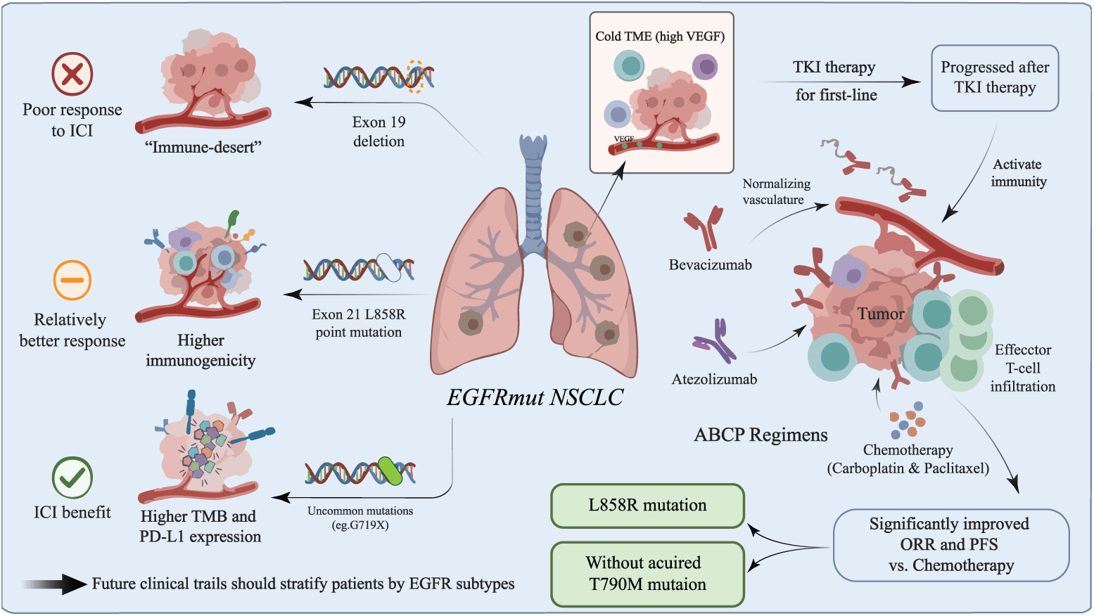
Figure 3. Heterogeneity of EGFR-mutant NSCLC and the rationale for combination therapy. ICI monotherapy generally has limited activity after EGFR TKI failure, although immune phenotype may vary by EGFR subtype, smoking exposure, and co-mutations. The ABCP regimen showed activity in exploratory subgroup analyses, but the relative contribution of each component and the optimal post-TKI strategy remain uncertain. Abbreviations: ABCP, atezolizumab, bevacizumab, carboplatin, and paclitaxel; EGFR, epidermal growth factor receptor; ICI, immune checkpoint inhibitor; ORR, objective response rate; PD-L1, programmed death-ligand 1; PFS, progression-free survival; TKI, tyrosine kinase inhibitor; TMB, tumor mutational burden; TME, tumor microenvironment; VEGF, vascular endothelial growth factor.
KRAS Mutations and Co-Mutations
Common KRAS substitutions include G12C, G12D, G12V, and G12A. KRAS-mutant NSCLC is frequently smoking-associated and may have higher TMB and an inflamed TME, but response cannot be inferred from KRAS status alone.52,53 Some studies report higher PD-L1 expression and TMB in G12C than in non-G12C tumors without a consistent difference in ICI efficacy, whereas preclinical and translational data suggest that G12D may be associated with lower PD-L1 expression and reduced CD8+ T-cell infiltration.52,54Clinical interpretation should therefore incorporate co-mutations and treatment context because the immune phenotype of KRAS-mutant tumors is strongly influenced by the co-mutation landscape (Fig. 4).
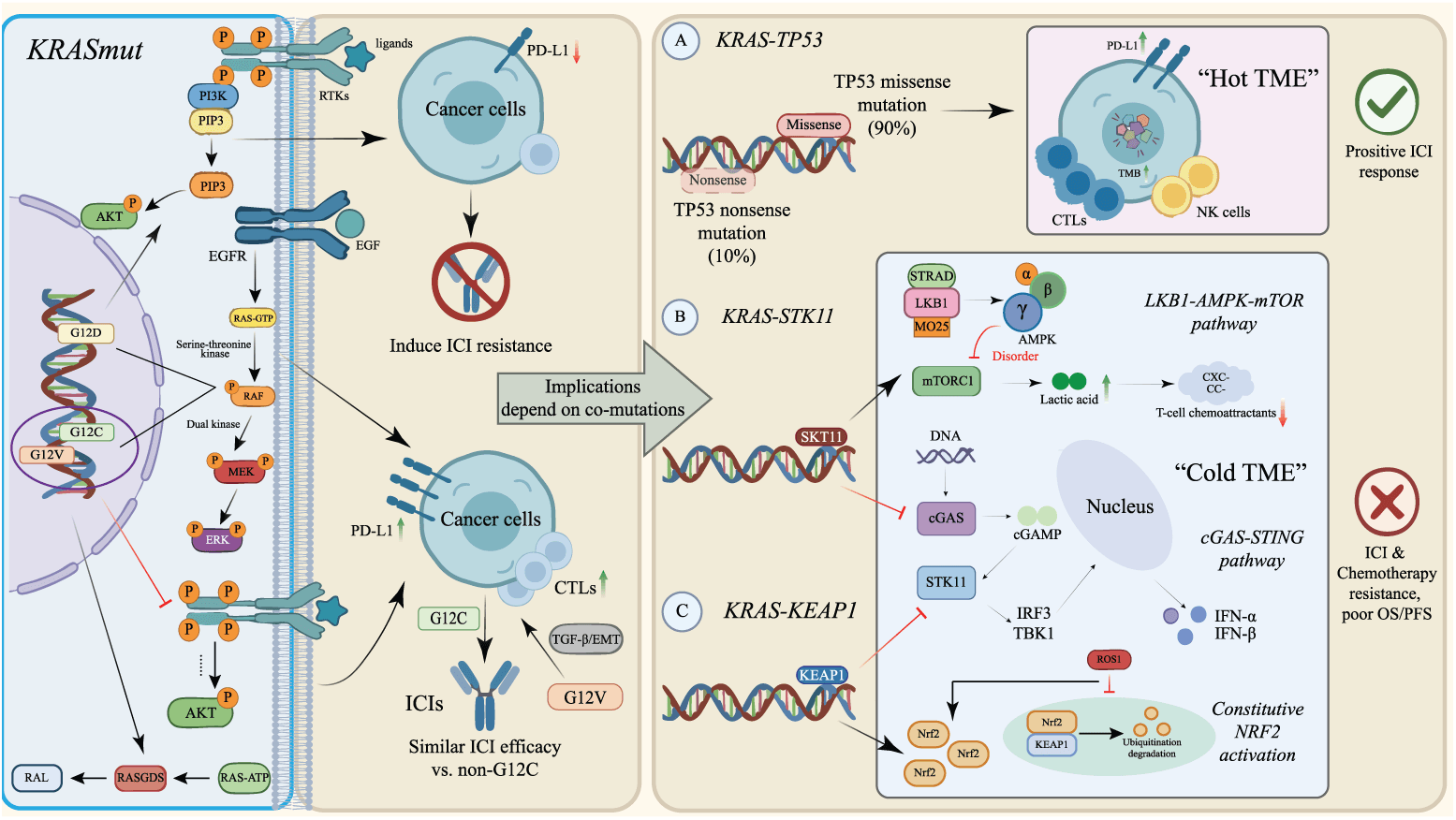
Figure 4. Heterogeneity of KRAS-mutant NSCLC and the influence of co-mutations on ICI outcomes. KRAS-TP53 co-mutation may be associated with a more inflamed phenotype in some cohorts, whereas STK11/LKB1 and KEAP1 alterations are associated with immune exclusion and adverse outcomes. These associations are not validated as standalone treatment-selection biomarkers. Abbreviations: AKT/PKB, protein kinase B; AMPK, AMP-activated protein kinase; ATP, adenosine triphosphate; cGAMP, 2′3′-cyclic GMP-AMP; cGAS, cyclic GMP-AMP synthase; CTL, cytotoxic T lymphocyte; EGF, epidermal growth factor; EGFR, epidermal growth factor receptor; EMT, epithelial–mesenchymal transition; ERK, extracellular signal-regulated kinase; GTP, guanosine triphosphate; ICI, immune checkpoint inhibitor; IFN, interferon; IRF3, interferon regulatory factor 3; KEAP1, kelch-like ECH-associated protein 1; KRAS, KRAS proto-oncogene, GTPase; MEK, mitogen-activated protein kinase kinase; MO25, mouse protein 25; mTORC, mechanistic target of rapamycin complex; NRF2, nuclear factor erythroid 2-related factor 2; NK, natural killer; OS, overall survival; PD-L1, programmed death-ligand 1; PFS, progression-free survival; PI3K, phosphatidylinositol 3-kinase; RAF, rapidly accelerated fibrosarcoma kinase; RAL, RAS-like protein; RAS, rat sarcoma virus family; RASGDS, RAS guanine nucleotide dissociation stimulator; ROS1, ROS proto-oncogene 1, receptor tyrosine kinase; RTK, receptor tyrosine kinase; STING, stimulator of interferon genes; STK11, serine/threonine kinase 11; STRAD, STE20-related kinase adaptor; TBK1, TANK-binding kinase 1; TMB, tumor mutational burden; TME, tumor microenvironment; TGF, transforming growth factor; TP53, tumor protein p53.
The interaction of KRAS with serine/threonine kinase 11 (STK11, also known as LKB1) or kelch-like ECH-associated protein 1 (KEAP1) is supported by mechanistic and clinical observations. In experimental models, STK11 loss can impair AMP-activated protein kinase (AMPK)-related metabolic control and suppress cGAS-STING/type I interferon signaling, reducing T-cell-recruiting chemokines and promoting an immune-excluded or immune-desert phenotype.55–57 These mechanistic data are biologically compelling, but they should not be equated with a prospectively validated clinical test. Retrospective cohorts associate STK11 alterations with poorer outcomes after immunotherapy and chemotherapy, indicating that at least part of the effect may be prognostic rather than uniquely predictive of PD-(L)1 resistance.58–61
KEAP1 loss activates NRF2-dependent antioxidant and metabolic programs and may reduce interferon signaling and immune-cell recruitment in preclinical systems.62,63 Clinical series consistently associate KEAP1, particularly with concurrent STK11 and KRAS alterations, with adverse prognosis. However, assay definitions, variant functional status, co-mutation structure, and treatment selection differ across cohorts. STK11 and KEAP1 should therefore be regarded as important risk stratifiers and investigational predictive markers, not as standalone validated reasons to withhold an otherwise indicated ICI regimen.
TP53 alterations are similarly heterogeneous. Some missense alterations and KRAS-TP53 co-mutations are associated with greater genomic instability, inflammatory signaling, and improved ICI outcomes in retrospective analyses, whereas truncating or functionally distinct variants may not confer the same phenotype.64,65 Mutation class, clonality, co-mutations, and tumor context should be considered, and TP53 status alone is not a validated treatment-selection biomarker.
Other Actionable Oncogenic Drivers
ICI outcomes also vary across tumors harboring alterations in anaplastic lymphoma kinase (ALK), ROS proto-oncogene 1, receptor tyrosine kinase (ROS1), RET proto-oncogene (RET), MET proto-oncogene, receptor tyrosine kinase (MET) exon 14 skipping, erb-b2 receptor tyrosine kinase 2 (ERBB2/HER2), and B-Raf proto-oncogene, serine/threonine kinase (BRAF). Rearrangement-driven tumors such as ALK-, ROS1-, and many RET-positive cancers commonly arise in never-smokers, have relatively low TMB, and show low response rates to ICI monotherapy despite occasional high PD-L1 expression. Outcomes in MET exon 14-skipping, ERBB2/HER2-mutant, and BRAF-altered NSCLC are more variable and may depend on smoking history, mutation subtype, and co-mutations.13,46 Matched targeted therapies should be prioritized when available; subsequent immunotherapy decisions should be individualized rather than based on a single driver category.
Combination Therapy and Treatment Modalities
PD-L1 expression, oncogenic-driver status, histology, disease burden, and patient fitness guide the selection of monotherapy or combination treatment in NSCLC. Chemotherapy, radiotherapy, antiangiogenic agents, dual checkpoint blockade, and other immunomodulatory strategies can broaden benefit, but efficacy must be balanced against toxicity, treatment discontinuation, quality of life, financial burden, and the possibility of exposing patients to unnecessary therapy (Fig. 5).
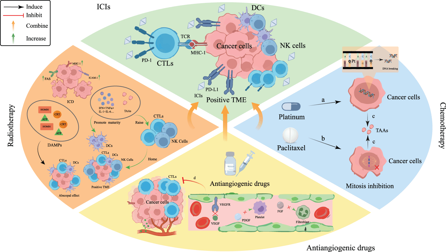
Figure 5. Mechanistic rationale for combining PD-(L)1 blockade with chemotherapy, radiotherapy, or antiangiogenic therapy. Cytotoxic therapies may induce DNA damage, mitotic disruption, antigen release, and immunogenic cell death, whereas antiangiogenic therapy may normalize vasculature and reduce VEGF-mediated immune suppression. These effects are context dependent and do not guarantee clinical synergy. Abbreviations: CRT, calreticulin; CTL, cytotoxic T lymphocyte; DAMP, damage-associated molecular pattern; DC, dendritic cell; FGF, fibroblast growth factor; HMGB1, high-mobility group box 1; ICAM, intercellular adhesion molecule; ICD, immunogenic cell death; ICI, immune checkpoint inhibitor; IFN, interferon; IL, interleukin; MDSC, myeloid-derived suppressor cell; MHC, major histocompatibility complex; NK, natural killer; PDGF, platelet-derived growth factor; PD-L1, programmed death-ligand 1; TAA, tumor-associated antigen; TAN, tumor-associated neutrophil; TCR, T-cell receptor; TME, tumor microenvironment; TNF, tumor necrosis factor; Treg, regulatory T cell; VEGF, vascular endothelial growth factor; VEGFR, vascular endothelial growth factor receptor.
Combination with Chemotherapeutic Agents
Chemoimmunotherapy improves response and survival in multiple first-line NSCLC settings and can benefit patients across PD-L1 categories.66 Reported ORRs of approximately 33%–47% with combination regimens versus approximately 20%–25% with historical monotherapy illustrate broader activity, but these figures are not directly comparable across trials.67,68 Chemoimmunotherapy does not eliminate immune-related adverse events (irAEs) and adds chemotherapy-related hematologic and nonhematologic toxicity.69 Associations between irAEs and longer progression-free or overall survival have been reported, but these analyses are vulnerable to guarantee-time and immortal-time bias and should not be interpreted as evidence that irAEs are beneficial.70,71 Randomized patient-reported outcome data from KEYNOTE-189 showed maintenance of global health status and delayed symptom deterioration in the trial population, although individual quality-of-life effects depend on baseline symptoms, comorbidity, cumulative chemotherapy, and toxicity.72 Financial burden and access may also influence the real-world value of prolonged combination treatment. Clinical selection is therefore essential, particularly for frail patients, those with cachexia, and those with high PD-L1 expression who may be candidates for monotherapy.73
Perioperative chemoimmunotherapy has improved pathological response and event-free survival in resectable NSCLC, including PD-L1-negative subgroups in pooled analyses.74 However, pathological response does not fully capture long-term benefit, and perioperative treatment can delay surgery or produce immune-related and chemotherapy-related toxicity. Future studies should define which patients require neoadjuvant plus adjuvant therapy, identify optimal chemotherapy backbones and treatment duration, and incorporate molecular residual disease and patient-reported outcomes.
Combination with Radiotherapy
Radiotherapy can induce immunogenic cell death, antigen release, and local immune remodeling, providing a rationale for combination with PD-(L)1 blockade.75,76 Combining stereotactic body radiotherapy (SBRT) with PD-(L)1 blockade can enhance local immune activation and has produced signals of increased out-of-field response in selected studies; however, a reproducible increase in the clinical abscopal effect has not been established.77–80 Proposed mechanisms include antigen release, altered tumor PD-L1 expression, increased CD8+ T-cell recruitment, and modulation of MDSCs and Tregs. Important unresolved variables include total dose, fraction size, irradiated site and volume, number of lesions, sequencing relative to ICI, treatment interval, baseline immune phenotype, and patient selection. High-dose SBRT may enhance antigen release but can also deplete radiosensitive lymphocytes, whereas low-dose radiation may alter stromal and myeloid compartments without reliably controlling tumor. Pulmonary toxicity is particularly relevant in NSCLC, and the interaction between radiation pneumonitis and immune-mediated pneumonitis requires careful prospective evaluation.81,82
Combination with Antiangiogenic Drugs
Antiangiogenic therapy may improve perfusion, reduce VEGF-mediated immune suppression, and facilitate lymphocyte trafficking, providing a rationale for combination with PD-(L)1 blockade. IMpower150 and ORIENT-31 support selected chemotherapy–antiangiogenic–immunotherapy regimens after targeted-therapy failure in EGFR-mutant NSCLC.50,51 In previously untreated, driver-negative, PD-L1-positive advanced NSCLC, the phase III HARMONi-2 study conducted in China showed longer progression-free survival with the PD-1/VEGF bispecific antibody ivonescimab than with pembrolizumab; however, overall survival maturity, external generalizability, regulatory status, and VEGF-related toxicity require consideration.83–85 Bleeding, hypertension, proteinuria, thromboembolic risk, regimen complexity, and cost must also be considered. Evidence from single-arm phase II studies, including endostatin-containing regimens, should not be interpreted as equivalent to randomized phase III confirmation.86
Combination with Other Immunotherapeutic Agents
Dual PD-(L)1 and CTLA-4 blockade can produce durable survival benefit and offers a chemotherapy-sparing option for selected patients, but it increases immune-mediated toxicity and requires careful patient selection.87,88 Neoadjuvant nivolumab plus relatlimab and pembrolizumab followed by delayed administration of the selective JAK1 inhibitor itacitinib have shown early clinical activity, but these approaches remain investigational in NSCLC.89,90 T-cell immunoreceptor with immunoglobulin and immunoreceptor tyrosine-based inhibitory motif domains (TIGIT) and other co-inhibitory receptors are biologically attractive targets; however, early-phase signals have not established a consistent new standard, and randomized confirmation is required.91
Antibody–Drug Conjugate Combinations
Antibody–drug conjugates (ADCs) deliver cytotoxic payloads to antigen-expressing tumor cells and may complement PD-(L)1 blockade through tumor-cell killing, antigen release, immunogenic cell death, and potential Fc-mediated immune effects. In the phase Ib TROPION-Lung02 study, datopotamab deruxtecan plus pembrolizumab, with or without platinum-based chemotherapy, demonstrated encouraging antitumor activity in previously untreated advanced NSCLC without actionable genomic alterations.92 The regimen remains investigational; the contribution of immune remodeling to clinical efficacy is incompletely established, and target expression, payload toxicity, stomatitis, hematologic toxicity, and interstitial lung disease/pneumonitis require careful evaluation. ADC–ICI combinations should therefore be described as promising but not uniformly proven across targets or settings.
Selected Phase III Studies and Unresolved Questions
At the May 2026 literature cutoff, TROPION-Lung08 (NCT05215340) was evaluating first-line datopotamab deruxtecan plus pembrolizumab versus pembrolizumab alone in advanced nonsquamous NSCLC with PD-L1 TPS ≥50%.93 PACIFIC-4/RTOG-3515 (NCT03833154) was evaluating durvalumab versus placebo after SBRT in unresected, node-negative early-stage NSCLC. Neither trial had reported definitive phase III efficacy results. These studies are designed to clarify incremental survival benefit, toxicity, quality of life, and the biomarker-defined populations most likely to benefit.
Individual Patient Characteristics
Host characteristics and concomitant medications can modify ICI outcomes through immune competence, systemic inflammation, pharmacokinetics, comorbidity, and treatment tolerance. Most supporting data are observational and are vulnerable to confounding; these variables should usually be interpreted as prognostic or hypothesis-generating rather than as independent treatment-selection biomarkers (Fig. 6).
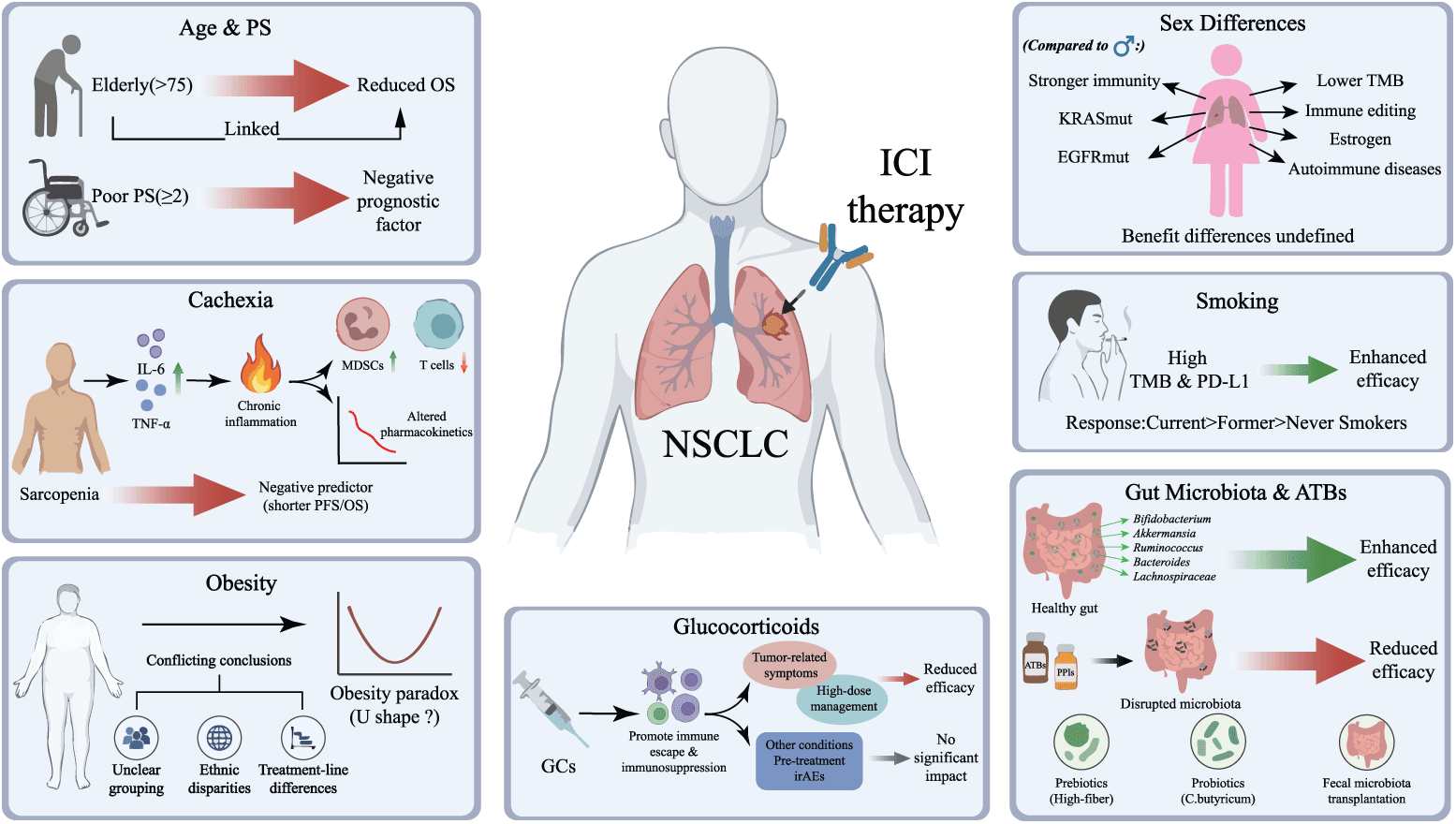
Figure 6. Host characteristics and concomitant exposures associated with outcomes during ICI therapy. Poor performance status, cachexia, sarcopenia, and baseline corticosteroid use for cancer-related symptoms are generally associated with inferior outcomes. Smoking history, obesity, microbiota composition, and antibiotic or proton-pump inhibitor exposure show context-dependent associations and should not be interpreted as causal or as independent treatment-selection criteria. Abbreviations: ATB, antibiotic; BMI, body mass index; EGFR, epidermal growth factor receptor; GC, glucocorticoid; ICI, immune checkpoint inhibitor; IL, interleukin; irAE, immune-related adverse event; KRAS, KRAS proto-oncogene, GTPase; MDSC, myeloid-derived suppressor cell; NSCLC, non-small cell lung cancer; OS, overall survival; PD-L1, programmed death-ligand 1; PFS, progression-free survival; PPI, proton-pump inhibitor; PS, performance status; TMB, tumor mutational burden; TNF, tumor necrosis factor.
Age and Performance Status
The effect of chronological age on ICI efficacy remains uncertain. Large observational cohorts and meta-analyses suggest that the magnitude of benefit may be attenuated in some patients aged 75 years or older, but older adults are underrepresented in randomized trials and differ substantially in frailty, comorbidity, organ function, performance status, and competing mortality.94,95 Ageing can increase mutation burden while simultaneously altering immune function; chronological age alone therefore provides an incomplete estimate of likely benefit.
Poorer performance status is one plausible explanation for less favorable outcomes in some older cohorts, but this is a potential confounding pathway rather than an established age-specific mechanism. ECOG performance status is a strong prognostic factor in advanced NSCLC, and patients with ECOG PS ≥2 generally have poorer outcomes than those with PS 0–1.96 Because poor PS may result from reversible tumor burden, chronic comorbidity, frailty, or terminal decline, its meaning should be assessed clinically rather than used as an isolated contraindication.
Cancer Cachexia
Cancer cachexia is a multifactorial syndrome characterized by progressive skeletal muscle loss, with or without adipose tissue loss, and is commonly accompanied by systemic inflammation. Cachexia and sarcopenia are consistently associated with shorter progression-free and overall survival during ICI-based treatment, although the extent to which they are predictive rather than prognostic remains uncertain.97–100 IL-6, TNF-α, myeloid-cell expansion, impaired T-cell function, altered drug clearance, reduced physiologic reserve, and treatment intolerance may contribute. Baseline nutritional and body-composition assessment is therefore clinically relevant, but interventional evidence showing that reversal of cachexia improves ICI efficacy is limited.
Obesity
Retrospective studies have reported an “obesity paradox,” in which overweight patients appear to have improved outcomes with ICIs, but results are inconsistent after adjustment for confounding.101–105 BMI cannot distinguish skeletal muscle from adipose tissue or characterize visceral versus subcutaneous adiposity. Sarcopenia, low muscle attenuation, visceral adiposity, and combined phenotypes such as sarcopenic obesity may be more biologically and prognostically informative than BMI alone.100 Future studies should use standardized CT-based body-composition measures, prespecified sex- and population-specific thresholds, and adjustment for performance status, smoking, disease burden, cachexia, and treatment line. BMI alone should not guide the selection of PD-(L)1 therapy.
Sex Differences
Sex-related differences in immunity, hormones, tobacco exposure, tumor genomics, and adverse-event risk provide a biological rationale for differential ICI outcomes.106–109 However, clinical studies and meta-analyses have produced inconsistent estimates, and observed differences may reflect histology, smoking history, EGFR/KRAS distribution, treatment regimen, or other confounders.110–113 Some analyses suggest greater relative benefit from chemoimmunotherapy than monotherapy in women, whereas others show no significant sex interaction. Current evidence does not support selecting or withholding PD-(L)1 therapy solely on the basis of sex; sex should instead be incorporated as a prespecified variable in adequately powered trials and multiparameter prediction models.
Smoking History
A history of tobacco exposure is associated with higher mutation burden and may enrich for response to ICI monotherapy in NSCLC, particularly in driver-negative disease.114,115 This association reflects smoking-related mutagenesis and correlated tumor biology; it does not imply that smoking is clinically desirable or should be continued. Tobacco use causes cancer, cardiovascular and pulmonary disease, treatment complications, and mortality. Smoking cessation should be recommended at every stage of care, and smoking history should be interpreted with PD-L1, oncogenic drivers, TMB, and other clinical factors rather than as an independent therapeutic target.
Gut Microbiota and Antibiotics
The gut microbiome can influence systemic immunity through microbial metabolites, epithelial-barrier function, and the gut–lung axis. In NSCLC cohorts, microbial diversity and taxa including Akkermansia, Bifidobacterium, Alistipes, and selected Firmicutes have been associated with ICI outcomes.116–122 These findings are associations rather than proof of causation. Reported taxa are not fully consistent across cohorts, and abundance may reflect geography, diet, ethnicity, comorbidity, medication exposure, stool-collection procedures, sequencing platform, reference database, and bioinformatic pipeline.
Antibiotic or proton-pump inhibitor exposure has been associated with poorer outcomes in several observational studies and meta-analyses, particularly when exposure occurs near ICI initiation.123–127 Nevertheless, confounding by infection, frailty, hospitalization, disease severity, indication, and cumulative exposure is difficult to eliminate, and other analyses have not demonstrated a causal detrimental effect.128 Clinically indicated antibiotics should not be withheld, but unnecessary exposure and prolonged broad-spectrum treatment should be avoided according to antimicrobial-stewardship principles. Microbiome studies should prespecify exposure windows and dose, account for indication, and use standardized sampling and analytic methods.
Dietary modification, probiotics, and fecal microbiota transplantation are being investigated as microbiome-directed strategies.129,130 Evidence in NSCLC remains preliminary: probiotic data are largely retrospective or small, and fecal microbiota transplantation has not become standard practice. Candidate biomarkers such as plasma citrulline and blood or stool bacterial measurements require independent validation.131 Intervention trials should include rigorous donor and product characterization, infection surveillance, dietary control, longitudinal sampling, and harmonized sequencing and bioinformatic workflows before clinical implementation.
Glucocorticoids
Glucocorticoids can suppress T-cell receptor signaling, alter lymphocyte and myeloid-cell states, promote Treg or M2-like programs, and potentially disrupt TLSs or the microbiome.132–134 These mechanisms support biological concern, but dose, duration, timing, and indication are critical. Short courses used for chemotherapy premedication or appropriate treatment of immune-related adverse events should not be interpreted in the same manner as persistent baseline steroids for cancer-related symptoms.
Retrospective studies associate baseline systemic glucocorticoid use, commonly defined as ≥10 mg/day prednisone equivalent, and early high-dose exposure with poorer response and survival during ICI therapy.135–140 However, indication bias is substantial: patients receiving glucocorticoids for dyspnea, brain metastases, cancer pain, or other palliative indications often have greater disease burden, poorer performance status, and worse prognosis independent of the drug itself. Analyses separating palliative from nonpalliative indications suggest that part of the observed association is attributable to this clinical context.136 Glucocorticoids should therefore be used at the lowest effective dose for the shortest appropriate duration, without delaying clinically necessary treatment of immune-related toxicity or other serious conditions.
Summary and Future Prospects
The efficacy of PD-(L)1 inhibitors in NSCLC is governed by interactions among tumor genomics, immune contexture, host condition, treatment exposure, and time. PD-L1 is clinically implemented, whereas most other proposed markers are incompletely standardized or are primarily prognostic. The central clinical implication is therefore not to replace one imperfect biomarker with another, but to integrate complementary variables while recognizing the quality and context of the supporting evidence.
Key Determinants of Therapeutic Response
An integrated framework should combine analytically validated baseline variables with early on-treatment measurements. Baseline components may include PD-L1 TPS, actionable driver and co-mutation profiles, calibrated TMB, tumor burden and metastatic sites, immune-cell and gene-expression features, body composition, performance status, and relevant medication exposure. Spatial transcriptomics and single-cell sequencing can refine the interpretation of T-cell states, TLSs, myeloid programs, stromal barriers, and cell–cell interactions, but these technologies require simplification and standardization before routine use.12–14
Dynamic biomarkers may be more informative than a static pretreatment specimen. Early reductions in circulating tumor DNA, particularly when integrated with circulating immune-cell features or imaging response, have been associated with durable clinical benefit in NSCLC.45,141 Serial ctDNA may also help distinguish molecular response from radiographic ambiguity, but low-shedding tumors and isolated intracranial disease remain important limitations. Additional dynamic measures may include peripheral immune-cell states, inflammatory indices, radiomic changes, and repeated tissue sampling when clinically feasible.
Artificial-intelligence-assisted models may integrate clinical, genomic, pathology, radiology, and longitudinal data at a scale that is difficult to interpret manually. Radiomic approaches have shown that imaging features can approximate CD8+ infiltration and associate with response to PD-(L)1 blockade.142 However, AI models are vulnerable to overfitting, dataset shift, missing data, and hidden confounding. External validation, calibration, transparent reporting, and demonstration of clinical utility are required before deployment.
Controversies and Directions
Major unresolved issues include cross-platform TMB calibration; assay and cutoff harmonization for spatial immune biomarkers; the predictive versus prognostic roles of STK11, KEAP1, cachexia, and concomitant medications; optimal sequencing of targeted therapy and ICI-based combinations in oncogene-driven disease; and reproducible microbiome measurement. Future studies should avoid simplistic positive/negative labels and should prespecify treatment setting, specimen type, timing, analytic method, and clinically meaningful decision thresholds.
Two trial concepts may accelerate progress. First, a biomarker-adaptive platform trial could assign or reassign patients using an integrated baseline profile comprising tumor genomics, PD-L1, spatial immune architecture, ctDNA, and host factors, with treatment arms added or discontinued according to prospectively defined Bayesian decision rules. Second, a longitudinal co-clinical trial could obtain serial ctDNA, peripheral immune profiling, imaging, and paired tissue or spatial analyses to identify early resistance states and trigger protocol-defined treatment adaptation. These designs should include patient-reported outcomes, toxicity, cost-effectiveness, and equitable access. Ultimately, precision immuno-oncology will require prospectively validated, dynamic multiparameter models that improve patient outcomes beyond current clinical and PD-L1-based selection.
Acknowledgment
None.
Authors’ Contributions
L.L. drafted the manuscript. Z.Z., Z.G., L.W., X.Q., and S.Z. participated in the major revision. L.W., X.Q., and S.Z. supervised the project. All authors have read and approved the final manuscript.
Funding Sources
This study was supported by grants from the Liaoning Province Science and Technology Plan Project (grant no. 2024JH2/102500025) and the National Natural Science Foundation of China (grant no. 82103338).
Ethics Declaration
Not applicable.
Data Availability Statement
No new data were generated or analyzed in this narrative review.
Generative AI Statement
Generative AI and AI-assisted technologies were used solely for language editing and manuscript refinement. The authors reviewed and approved all changes and take full responsibility for the content.
Conflict-of-Interest Statement
The authors declare that the research was conducted in the absence of any commercial or financial relationships that could be construed as a potential conflict of interest.
References
[1] Reck M, Rodríguez-Abreu D, Robinson AG, et al. Pembrolizumab versus chemotherapy for PD-L1-positive non-small-cell lung cancer. N Engl J Med. November 10, 2016;375(19):1823–1833. doi:10.1056/NEJMoa1606774.
[2] Guo L, Liang J, Dai W, et al. PD-1/L1 with or without CTLA-4 inhibitors versus chemotherapy in advanced non-small cell lung cancer. Cancer Control. January–December 2022;29:10732748221107590. doi:10.1177/10732748221107590.
[3] Hu Y, Liu S, Wang L, Liu Y, Zhang D, Zhao Y. Treatment-free survival after discontinuation of immune checkpoint inhibitors in mNSCLC: a systematic review and meta-analysis. Front Immunol. 2023;14:1202822. doi:10.3389/fimmu.2023.1202822.
[4] Wu B, Zhang B, Li B, Wu H, Jiang M. Cold and hot tumors: from molecular mechanisms to targeted therapy. Signal Transduct Target Ther. October 18, 2024;9(1):274. doi:10.1038/s41392-024-01979-x.
[5] Zhang P, Wang D, Zhou G, et al. Novel post-translational modification learning signature reveals B4GALT2 as an immune exclusion regulator in lung adenocarcinoma. J Immunother Cancer. February 25, 2025;13(2):e010787. doi:10.1136/jitc-2024-010787.
[6] Restle D, Dux J, Li X, et al. Organ-specific heterogeneity in tumor-infiltrating immune cells and cancer antigen expression in primary and autologous metastatic lung adenocarcinoma. J Immunother Cancer. June 2023;11(6):e006609. doi:10.1136/jitc-2022-006609.
[7] Scuoppo C, Ramirez R, Leong SF, et al. The C/EBPβ antagonist peptide lucicebtide (ST101) induces macrophage polarization toward a pro-inflammatory phenotype and enhances anti-tumor immune responses. Front Immunol. 2025;16:1522699. doi:10.3389/fimmu.2025.1522699.
[8] Brummel K, Eerkens AL, de Bruyn M, Nijman HW. Tumour-infiltrating lymphocytes: from prognosis to treatment selection. Br J Cancer. February 2023;128(3):451–458. doi:10.1038/s41416-022-02119-4.
[9] Kamphorst AO, Wieland A, Nasti T, et al. Rescue of exhausted CD8 T cells by PD-1-targeted therapies is CD28-dependent. Science. March 31, 2017;355(6332):1423–1427. doi:10.1126/science.aaf0683.
[10] Fang Z, Ding X, Huang H, Jiang H, Jiang J, Zheng X. Revolutionizing tumor immunotherapy: unleashing the power of progenitor exhausted T cells. Cancer Biol Med. May 31, 2024;21(6):499–512. doi:10.20892/j.issn.2095-3941.2024.0105.
[11] Liu Z, Zhang Y, Ma N, et al. Progenitor-like exhausted SPRY1(+)CD8(+) T cells potentiate responsiveness to neoadjuvant PD-1 blockade in esophageal squamous cell carcinoma. Cancer Cell. November 13, 2023;41(11):1852–1870.e9. doi:10.1016/j.ccell.2023.09.011.
[12] Koh J, Kim S, Woo Y. TCF1+PD-1+ tumour-infiltrating lymphocytes predict a favorable response and prolonged survival after immune checkpoint inhibitor therapy for non-small-cell lung cancer. Eur J Cancer. 2022;174:10–20. doi:10.1016/j.ejca.2022.07.004.
[13] Sun X, Liu W, Sun L. Maturation and abundance of tertiary lymphoid structures are associated with the efficacy of neoadjuvant chemoimmunotherapy in resectable non-small cell lung cancer. J Immunother Cancer. 2022;10(11):e005531. doi:10.1136/jitc-2022-005531.
[14] Peyraud F, Guégan J, Rey C. Spatially resolved transcriptomics reveal the determinants of primary resistance to immunotherapy in NSCLC with mature tertiary lymphoid structures. Cell Rep Med. 2025;6(5):101934. doi:10.1016/j.xcrm.2025.101934.
[15] Cai S, Yang G, Hu M, et al. Spatial cell interplay networks of regulatory T cells predict recurrence in patients with operable non-small cell lung cancer. Cancer Immunol Immunother. August 2, 2024;73(10):189. doi:10.1007/s00262-024-03762-x.
[16] Liu Y, Han Y, Zhang Y, Lv T, Peng X, Huang J. LncRNAs has been identified as regulators of Myeloid-derived suppressor cells in lung cancer. Front Immunol. 2023;14:1067520. doi:10.3389/fimmu.2023.1067520.
[17] Chow A, Uddin FZ, Liu M, et al. The ectonucleotidase CD39 identifies tumor-reactive CD8(+) T cells predictive of immune checkpoint blockade efficacy in human lung cancer. Immunity. January 10, 2023;56(1):93–106.e6. doi:10.1016/j.immuni.2022.12.001.
[18] Lasser SA, Ozbay Kurt FG, Arkhypov I, Utikal J, Umansky V. Myeloid-derived suppressor cells in cancer and cancer therapy. Nat Rev Clin Oncol. February 2024;21(2):147–164. doi:10.1038/s41571-023-00846-y.
[19] Bronte G, Petracci E, De Matteis S, et al. High levels of circulating monocytic myeloid-derived suppressive-like cells are associated with the primary resistance to immune checkpoint inhibitors in advanced non-small cell lung cancer: an exploratory analysis. Front Immunol. 2022;13:866561. doi:10.3389/fimmu.2022.866561.
[20] Jeong H, Koh J, Kim S, et al. Cell-intrinsic PD-L1 signaling drives immunosuppression by myeloid-derived suppressor cells through IL-6/Jak/Stat3 in PD-L1-high lung cancer. J Immunother Cancer. March 6, 2025;13(3):e010612. doi:10.1136/jitc-2024-010612.
[21] Raniszewska A, Vroman H, Dumoulin D, Cornelissen R, Aerts J, Domagała-Kulawik J. PD-L1(+) lung cancer stem cells modify the metastatic lymph-node immunomicroenvironment in NSCLC patients. Cancer Immunol Immunother. February 2021;70(2):453–461. doi:10.1007/s00262-020-02648-y.
[22] Zhu P, Jin Z, Kang G, et al. Alpha5 nicotinic acetylcholine receptor mediated immune escape of lung adenocarcinoma via STAT3/Jab1-PD-L1 signalling. Cell Commun Signal. August 15, 2022;20(1):121. doi:10.1186/s12964-022-00934-z.
[23] Kim Y, Park K, Kim YJ, et al. Immunomodulation of HDAC inhibitor entinostat potentiates the anticancer effects of radiation and PD-1 blockade in the murine lewis lung carcinoma model. Int J Mol Sci. December 8, 2022;23(24):15539. doi:10.3390/ijms232415539.
[24] Liao S, Sun H, Lu H, et al. Neutrophil-to-lymphocyte ratio-based prognostic score can predict outcomes in patients with advanced non-small cell lung cancer treated with immunotherapy plus chemotherapy. BMC Cancer. April 15, 2025;25(1):697. doi:10.1186/s12885-025-13811-y.
[25] Longueville E, Dewolf M, Dalstein V, et al. Comparing neutrophil-to-lymphocyte ratio (NLR), absolute neutrophil count (ANC) and derived NLR as predictive biomarkers in first-line immunotherapy for non-small cell lung cancer: a retrospective study. Transl Lung Cancer Res. April 30, 2025;14(4):1212–1230. doi:10.21037/tlcr-24-808.
[26] Kashimura S, Sato M, Inagaki T, et al. Relationship between the combination of platelet count and neutrophil-lymphocyte ratio and prognosis of patients with advanced non-small cell lung cancer treated with immune checkpoint inhibitors plus chemotherapy: A retrospective cohort study. Thorac Cancer. October 2024;15(28):2049–2060. doi:10.1111/1759-7714.15437.
[27] Xue J, Liu C, Shao J, et al. Predictive value of neutrophil-to-lymphocyte ratio for immune checkpoint inhibitor-related myocarditis among patients treated for non-small-cell lung cancer. Cancer Innov. April 2025;4(2):e163. doi:10.1002/cai2.163.
[28] Kim H, Chung JH. PD-L1 testing in non-small cell lung cancer: past, present, and future. J Pathol Transl Med. July 2019;53(4):199–206. doi:10.4132/jptm.2019.04.24.
[29] Rittmeyer A, Barlesi F, Waterkamp D, et al. Atezolizumab versus docetaxel in patients with previously treated non-small-cell lung cancer (OAK): a phase 3, open-label, multicentre randomised controlled trial. Lancet. January 21, 2017;389(10066):255–265. doi:10.1016/s0140-6736(16)32517-x.
[30] Tsao M, Kerr K, Kockx M. PD-L1 immunohistochemistry comparability study in real-life clinical samples: results of blueprint phase 2 project. J Thorac Oncol. 2018;13(9):1302–1311. doi:10.1016/j.jtho.2018.05.013.
[31] Kowanetz M, Zou W, Gettinger SN, et al. Differential regulation of PD-L1 expression by immune and tumor cells in NSCLC and the response to treatment with atezolizumab (anti-PD-L1). Proc Natl Acad Sci USA. October 23, 2018;115(43):E10119–e10126. doi:10.1073/pnas.1802166115.
[32] Schumacher TN, Scheper W, Kvistborg P. Cancer neoantigens. Annu Rev Immunol. April 26, 2019;37:173–200. doi:10.1146/annurev-immunol-042617-053402.
[33] Meri-Abad M, Moreno-Manuel A, García SG, et al. Clinical and technical insights of tumour mutational burden in non-small cell lung cancer. Crit Rev Oncol Hematol. February 2023;182:103891. doi:10.1016/j.critrevonc.2022.103891.
[34] Di Federico A, Alden SL, Smithy JW, et al. Intrapatient variation in PD-L1 expression and tumor mutational burden and the impact on outcomes to immune checkpoint inhibitor therapy in patients with non-small-cell lung cancer. Ann Oncol. October 2024;35(10):902–913. doi:10.1016/j.annonc.2024.06.014.
[35] Rizvi H, Sanchez-Vega F, La K, et al. Molecular determinants of response to anti-programmed cell death (PD)-1 and anti-programmed death-ligand 1 (PD-L1) blockade in patients with non-small-cell lung cancer profiled with targeted next-generation sequencing. J Clin Oncol. March 1, 2018;36(7):633–641. doi:10.1200/jco.2017.75.3384.
[36] Kowanetz M, Zou W, Shames D, et al. OA20.01 Tumor Mutation Burden (TMB) is associated with improved efficacy of atezolizumab in 1L and 2L+ NSCLC patients. Journal of Thoracic Oncology. 2017;12(1):S321–S322. doi:10.1016/j.jtho.2016.11.343.
[37] Carbone DP, Reck M, Paz-Ares L, et al. First-line nivolumab in stage IV or recurrent non-small-cell lung cancer. N Engl J Med. June 22, 2017;376(25):2415–2426. doi:10.1056/NEJMoa1613493.
[38] Hellmann MD, Ciuleanu TE, Pluzanski A, et al. Nivolumab plus ipilimumab in lung cancer with a high tumor mutational burden. N Engl J Med. May 31, 2018;378(22):2093–2104. doi:10.1056/NEJMoa1801946.
[39] Zhao P, Li L, Jiang X, Li Q. Mismatch repair deficiency/microsatellite instability-high as a predictor for anti-PD-1/PD-L1 immunotherapy efficacy. J Hematol Oncol. May 31, 2019;12(1):54. doi:10.1186/s13045-019-0738-1.
[40] Palmeri M, Mehnert J, Silk AW, et al. Real-world application of tumor mutational burden-high (TMB-high) and microsatellite instability (MSI) confirms their utility as immunotherapy biomarkers. ESMO Open. February 2022;7(1):100336. doi:10.1016/j.esmoop.2021.100336.
[41] Stenzinger A, Allen JD, Maas J, et al. Tumor mutational burden standardization initiatives: recommendations for consistent tumor mutational burden assessment in clinical samples to guide immunotherapy treatment decisions. Genes Chromosomes Cancer. August 2019;58(8):578–588. doi:10.1002/gcc.22733.
[42] Ramos-Paradas J, Hernández-Prieto S, Lora D, et al. Tumor mutational burden assessment in non-small-cell lung cancer samples: results from the TMB(2) harmonization project comparing three NGS panels. J Immunother Cancer. May 2021;9(5):e001904. doi:10.1136/jitc-2020-001904.
[43] Furtado LV, Bifulco C, Dolderer D, et al. Recommendations for tumor mutational burden assay validation and reporting: a joint consensus recommendation of the association for Molecular Pathology, College of American Pathologists, and Society for Immunotherapy of Cancer. J Mol Diagn. August 2024;26(8):653–668. doi:10.1016/j.jmoldx.2024.05.002.
[44] Yarchoan M, Albacker L, Hopkins A. PD-L1 expression and tumor mutational burden are independent biomarkers in most cancers. Journal of Clinical Investigation Insight. 2019;4(6):e126908. doi:10.1172/jci.insight.126908.
[45] Nabet B, Esfahani M, Moding E. Noninvasive early identification of therapeutic benefit from immune checkpoint inhibition. Cell. 2020;183(2):363–376.e13. doi:10.1016/j.cell.2020.09.001.
[46] Mazieres J, Drilon A, Lusque A. Immune checkpoint inhibitors for patients with advanced lung cancer and oncogenic driver alterations: results from the IMMUNOTARGET registry. Ann Oncol. 2019;30(8):1321–1328. doi:10.1093/annonc/mdz167.
[47] Chen Z, Pang L, Yang Y, et al. Immunotherapy efficacy between exon 19 deletion and exon 21 L858R mutation in advanced EGFR mutant non-small-cell lung cancer: a direct and indirect meta-analysis. Transl Lung Cancer Res. February 28, 2025;14(2):422–430. doi:10.21037/tlcr-24-884.
[48] Tian J, Shi Z, Zhao L, et al. Revolutionizing NSCLC treatment: immunotherapy strategies for EGFR-TKIs resistance. Clin Respir J. December 2024;18(12):e70037. doi:10.1111/crj.70037.
[49] Ma T, Jiao J, Huo R, et al. PD-L1 expression, tumor mutational burden, and immune cell infiltration in non-small cell lung cancer patients with epithelial growth factor receptor mutations. Front Oncol. 2022;12:922899. doi:10.3389/fonc.2022.922899.
[50] Reck M, Mok TSK, Nishio M, et al. Atezolizumab plus bevacizumab and chemotherapy in non-small-cell lung cancer (IMpower150): key subgroup analyses of patients with EGFR mutations or baseline liver metastases in a randomised, open-label phase 3 trial. Lancet Respir Med. May 2019;7(5):387–401. doi:10.1016/s2213-2600(19)30084-0.
[51] Lu S, Wu L, Jian H, et al. Sintilimab plus bevacizumab biosimilar IBI305 and chemotherapy for patients with EGFR-mutated non-squamous non-small-cell lung cancer who progressed on EGFR tyrosine-kinase inhibitor therapy (ORIENT-31): first interim results from a randomised, double-blind, multicentre, phase 3 trial. Lancet Oncol. September 2022;23(9):1167–1179. doi:10.1016/s1470-2045(22)00382-5.
[52] Frost MG, Jensen KJ, Gotfredsen DR, et al. KRAS G12C mutated advanced non-small cell lung cancer (NSCLC): characteristics, treatment patterns and overall survival from a Danish nationwide observational register study. Lung Cancer. April 2023;178:172–182. doi:10.1016/j.lungcan.2023.02.021.
[53] Singhal A, Li BT, O’Reilly EM. Targeting KRAS in cancer. Nat Med. April 2024;30(4):969–983. doi:10.1038/s41591-024-02903-0.
[54] Wang M, Zhu L, Yang X, Li J, Liu Y, Tang Y. Targeting immune cell types of tumor microenvironment to overcome resistance to PD-1/PD-L1 blockade in lung cancer. Front Pharmacol. 2023;14:1132158. doi:10.3389/fphar.2023.1132158.
[55] Goldschmid H, Kluck K, Ball M, et al. Spatial profiling of the microenvironment reveals low intratumoral heterogeneity and STK11-associated immune evasion in therapy-naïve lung adenocarcinomas. Lung Cancer. June 2023;180:107212. doi:10.1016/j.lungcan.2023.107212.
[56] Lenahan SM, Sarausky HM, Deming P, Seward DJ. STK11 loss leads to YAP1-mediated transcriptional activation in human KRAS-driven lung adenocarcinoma cell lines. Cancer Gene Ther. January 2024;31(1):1–8. doi:10.1038/s41417-023-00687-y.
[57] Zheng J, Deng Y, Huang B, Chen X. Prognostic implications of STK11 with different mutation status and its relationship with tumor-infiltrating immune cells in non-small cell lung cancer. Front Immunol. 2024;15:1387896. doi:10.3389/fimmu.2024.1387896.
[58] Skoulidis F, Goldberg ME, Greenawalt DM, et al. STK11/LKB1 mutations and PD-1 inhibitor resistance in KRAS-mutant lung adenocarcinoma. Cancer Discov. July 2018;8(7):822–835. doi:10.1158/2159-8290.Cd-18-0099.
[59] Ricciuti B, Lamberti G, Puchala SR, et al. Genomic and immunophenotypic landscape of acquired resistance to PD-(L)1 blockade in non-small-cell lung cancer. J Clin Oncol. April 10, 2024;42(11):1311–1321. doi:10.1200/jco.23.00580.
[60] Papillon-Cavanagh S, Doshi P, Dobrin R, Szustakowski J, Walsh AM. STK11 and KEAP1 mutations as prognostic biomarkers in an observational real-world lung adenocarcinoma cohort. ESMO Open. April 2020;5(2):e000706. doi:10.1136/esmoopen-2020-000706.
[61] Liang Y, Maeda O, Kondo C, Nishida K, Ando Y. Effects of KRAS, STK11, KEAP1, and TP53 mutations on the clinical outcomes of immune checkpoint inhibitors among patients with lung adenocarcinoma. PLoS One. 2024;19(7):e0307580. doi:10.1371/journal.pone.0307580.
[62] Xu K, Ma J, Hall SRR, Peng RW, Yang H, Yao F. Battles against aberrant KEAP1-NRF2 signaling in lung cancer: intertwined metabolic and immune networks. Theranostics. 2023;13(2):704–723. doi:10.7150/thno.80184.
[63] Shrestha A, Li Y, Huang L, et al. Redox phenotype confers T cell-exclusion microenvironment and resistance to immunotherapy by suppressing STING/MDA5 expression and interferon signaling in lung cancers harboring KEAP1/STK11 mutations. Front Oncol. 2025;15:1676797. doi:10.3389/fonc.2025.1676797.
[64] Mathiot L, Nigen B, Goronflot T, et al. Prognostic impact of TP53 mutations in metastatic nonsquamous non-small-cell lung cancer. Clin Lung Cancer. May 2024;25(3):244–253.e2. doi:10.1016/j.cllc.2023.12.004.
[65] Sun H, Liu SY, Zhou JY, et al. Specific TP53 subtype as biomarker for immune checkpoint inhibitors in lung adenocarcinoma. EBioMedicine. October 2020;60:102990. doi:10.1016/j.ebiom.2020.102990.
[66] Wang Y, Han H, Zhang F, et al. Immune checkpoint inhibitors alone vs immune checkpoint inhibitors-combined chemotherapy for NSCLC patients with high PD-L1 expression: a network meta-analysis. Br J Cancer. September 2022;127(5):948–956. doi:10.1038/s41416-022-01832-4.
[67] Lahiri A, Maji A, Potdar PD, et al. Lung cancer immunotherapy: progress, pitfalls, and promises. Mol Cancer. February 21, 2023;22(1):40. doi:10.1186/s12943-023-01740-y.
[68] Gettinger S, Rizvi NA, Chow LQ, et al. Nivolumab monotherapy for first-line treatment of advanced non-small-cell lung cancer. J Clin Oncol. September 1, 2016;34(25):2980–2987. doi:10.1200/jco.2016.66.9929.
[69] Uematsu M, Tsukita Y, Tozuka T, et al. First-line immune checkpoint inhibitors alone or in combination with chemotherapy in real-life elderly patients with advanced non-small cell lung cancer (NEJ057). J Clin Oncol. 2023;41(16_suppl):9012. doi:10.1200/JCO.2023.41.16_suppl.9012.
[70] Serino M, Freitas C, Martins M, et al. Predictors of immune-related adverse events and outcomes in patients with NSCLC treated with immune-checkpoint inhibitors. Pulmonology. July–August 2024;30(4):352–361. doi:10.1016/j.pulmoe.2022.03.003.
[71] Heyward J, Lesko CR, Murray JC, Mehta HB, Segal JB. Harm-benefit balance of immune checkpoint inhibitors in non-small cell lung cancer. JAMA Oncol. July 1, 2025;11(7):707–716. doi:10.1001/jamaoncol.2025.0985.
[72] Garassino M, Gadgeel S, Esteban E. Patient-reported outcomes following pembrolizumab or placebo plus pemetrexed and platinum in patients with previously untreated, metastatic, non-squamous non-small-cell lung cancer (KEYNOTE-189). Lancet Oncol. 2020;21(3):387–397. doi:10.1016/s1470-2045(19)30801-0.
[73] Kawachi H, Yamada T, Tamiya M, et al. Clinical impact of cancer cachexia on the outcome of patients with non-small cell lung cancer with PD-L1 tumor proportion scores of ≥50% receiving pembrolizumab monotherapy versus immune checkpoint inhibitor with chemotherapy. Oncoimmunology. December 2025;14(1):2442116. doi:10.1080/2162402x.2024.2442116.
[74] Zhang SL, Tian Y, Yu J, et al. Is neoadjuvant immunotherapy necessary in patients with programmed death ligand 1 expression-negative resectable non-small cell lung cancer? A systematic review and meta-analysis. Lung Cancer. May 2024;191:107799. doi:10.1016/j.lungcan.2024.107799.
[75] Chinese expert consensus on the combination of immune checkpoint inhibitors and radiotherapy for lung cancer (2025 edition). Zhonghua Zhong Liu Za Zhi. 2025;47(1):39–64. doi:10.3760/cma.j.cn112152-20240929-00422.
[76] Hayashi K, Nikolos F, Lee YC, et al. Tipping the immunostimulatory and inhibitory DAMP balance to harness immunogenic cell death. Nat Commun. December 7, 2020;11(1):6299. doi:10.1038/s41467-020-19970-9.
[77] Yao D, Zhu X, Guo J, et al. A real-world analysis of stereotactic body radiotherapy combined with immunotherapy in advanced or recurrent non-small cell lung cancer (NSCLC): a single-center experience. Clin Transl Radiat Oncol. July 2024;47:100787. doi:10.1016/j.ctro.2024.100787.
[78] Tsur E, Blumenfeld P, Rottenberg Y, et al. Outcomes of extracranial stereotactic body radiation therapy for induced oligometastatic non-small cell lung cancer on novel systemic therapy. Transl Lung Cancer Res. March 29, 2024;13(3):465–474. doi:10.21037/tlcr-23-802.
[79] Theelen W, Peulen HMU, Lalezari F, et al. Effect of pembrolizumab after stereotactic body radiotherapy vs pembrolizumab alone on tumor response in patients with advanced non-small cell lung cancer: results of the PEMBRO-RT phase 2 randomized clinical trial. JAMA Oncol. September 1, 2019;5(9):1276–1282. doi:10.1001/jamaoncol.2019.1478.
[80] Welsh J, Menon H, Chen D, et al. Pembrolizumab with or without radiation therapy for metastatic non-small cell lung cancer: a randomized phase I/II trial. J Immunother Cancer. October 2020;8(2):e001001. doi:10.1136/jitc-2020-001001.
[81] Patel RB, Hernandez R, Carlson P, et al. Low-dose targeted radionuclide therapy renders immunologically cold tumors responsive to immune checkpoint blockade. Sci Transl Med. July 14, 2021;13(602):728. doi:10.1126/scitranslmed.abb3631.
[82] Zhou X, Zhou L, Yao Z, et al. Safety and tolerability of low-dose radiation and stereotactic body Radiotherapy + Sintilimab for treatment-Naïve Stage IV PD-L1+ non-small cell lung cancer patients. Clin Cancer Res. October 13, 2023;29(20):4098–4108. doi:10.1158/1078-0432.Ccr-23-0315.
[83] Huang L, Zhang S, Sun L, Ma J, Han C. Immune checkpoint inhibitors in epidermal growth factor receptor-tyrosine kinase inhibitor-resistant chemotherapy-Naïve advanced non-small cell lung cancer: a meta-analysis based on eight randomized trials. JCO Precis Oncol. July 2025;9:e2400907. doi:10.1200/po-24-00907.
[84] Zhao Y, Chen G, Chen J, et al. AK112, a novel PD-1/VEGF bispecific antibody, in combination with chemotherapy in patients with advanced non-small cell lung cancer (NSCLC): an open-label, multicenter, phase II trial. eClinicalMedicine. 2023;62:102106. doi:10.1016/j.eclinm.2023.102106.
[85] Tsukada A, Morita C, Shimizu Y, et al. Efficacy of first-line immune checkpoint inhibitor and anti-angiogenic agent combination therapy for Kirsten rat sarcoma viral antigen-mutant advanced non-small-cell lung cancer: a systematic review and network meta-analysis. Thorac Cancer. September 2024;15(25):1854–1862. doi:10.1111/1759-7714.15413.
[86] Wang D, Feng Y, Bai Y, et al. Three days of CIV rh-Endostatin in combination with PD-1 antibody plus chemotherapy as first-line regimen for EGFR/ALK-negative, advanced or metastatic, non-squamous non–small cell lung cancer (NSCLC): a open label, multicenter, phase II and cohort study (ENPOWER). J Clin Oncol. 2022;40(16_suppl):9031. doi:10.1200/JCO.2022.40.16_suppl.9031.
[87] Deng H, Zhou C. From CheckMate 227 to CheckMate 9LA: rethinking the status of chemotherapy in the immunotherapy era-chemo-free or chemo-reform? Transl Lung Cancer Res. April 2021;10(4):1924–1927. doi:10.21037/tlcr-21-179.
[88] Brahmer JR, Lee JS, Ciuleanu TE, et al. Five-year survival outcomes with nivolumab plus ipilimumab versus chemotherapy as first-line treatment for metastatic non-small-cell lung cancer in CheckMate 227. J Clin Oncol. February 20, 2023;41(6):1200–1212. doi:10.1200/jco.22.01503.
[89] Schuler M, Cuppens K, Plönes T, et al. Neoadjuvant nivolumab with or without relatlimab in resectable non-small-cell lung cancer: a randomized phase 2 trial. Nat Med. June 2024;30(6):1602–1611. doi:10.1038/s41591-024-02965-0.
[90] Mathew D, Marmarelis ME, Foley C, et al. Combined JAK inhibition and PD-1 immunotherapy for non-small cell lung cancer patients. Science. June 21, 2024;384(6702):eadf1329. doi:10.1126/science.adf1329.
[91] Rousseau A, Parisi C, Barlesi F. Anti-TIGIT therapies for solid tumors: a systematic review. ESMO Open. April 2023;8(2):101184. doi:10.1016/j.esmoop.2023.101184.
[92] Levy B, Paz-Ares L, Lin C-C, et al. Datopotamab deruxtecan plus pembrolizumab with or without platinum-based chemotherapy for advanced or metastatic NSCLC: the phase Ib TROPION-Lung02 trial. J Thorac Oncol. 2026;21(7):103688. doi:10.1016/j.jtho.2026.103688.
[93] Levy B, Felip E, Reck M. TROPION-Lung08: phase III study of datopotamab deruxtecan plus pembrolizumab as first-line therapy for advanced NSCLC. Future Oncol. 2023;19(21):1461–1472. doi:10.2217/fon-2023-0230.
[94] Voruganti T, Soulos PR, Mamtani R, Presley CJ, Gross CP. Association between age and survival trends in advanced non-small cell lung cancer after adoption of immunotherapy. JAMA Oncol. March 1, 2023;9(3):334–341. doi:10.1001/jamaoncol.2022.6901.
[95] Yao J, Li S, Bai L, et al. Efficacy and safety of immune checkpoint inhibitors in elderly patients with advanced non-small cell lung cancer: a systematic review and meta-analysis. EClinicalMedicine. March 2025;81:103081. doi:10.1016/j.eclinm.2025.103081.
[96] Zhu Y, Wu S, Ma F, Uemura T, Hu N, Wang C. Predictive factors for the efficacy of immune checkpoint inhibitors in advanced non-small cell lung cancer: a retrospective study. J Thorac Dis. June 30, 2025;17(6):4159–4176. doi:10.21037/jtd-2025-868.
[97] Morimoto K, Uchino J, Yokoi T, et al. Impact of cancer cachexia on the therapeutic outcome of combined chemoimmunotherapy in patients with non-small cell lung cancer: a retrospective study. Oncoimmunology. 2021;10(1):1950411. doi:10.1080/2162402x.2021.1950411.
[98] Guo Y, Remaily BC, Thomas J, et al. Antibody drug clearance: an underexplored marker of outcomes with checkpoint inhibitors. Clin Cancer Res. March 1, 2024;30(5):942–958. doi:10.1158/1078-0432.Ccr-23-1683.
[99] Yu Y, Yan L, Huang T, Wu Z, Liu J. Cancer cachexia reduces the efficacy of immune checkpoint inhibitors in cancer patients. Aging (Albany NY). March 11, 2024;11(6):5354–5369. doi:10.18632/aging.205652.
[100] Shiroyama T, Nagatomo I, Koyama S. Impact of sarcopenia in patients with advanced non-small cell lung cancer treated with PD-1 inhibitors: a preliminary retrospective study. Sci Rep. 2019;9:2447. doi:10.1038/s41598-019-39120-6.
[101] Sun Y, Dang Q, Ge Y, et al. Prognostic value of body mass index for first-line chemoimmunotherapy combinations in advanced non-small cell lung cancer in Chinese population. Heliyon. June 15, 2024;10(11):e31863. doi:10.1016/j.heliyon.2024.e31863.
[102] Ihara Y, Sawa K, Imai T, et al. Immunotherapy and overall survival among patients with advanced non-small cell lung cancer and obesity. JAMA Netw Open. August 1, 2024;7(8):e2425363. doi:10.1001/jamanetworkopen.2024.25363.
[103] Jain A, Zhang S, Shanley RM, et al. Nonlinear association between body mass index and overall survival in advanced NSCLC patients treated with immune checkpoint blockade. Cancer Immunol Immunother. May 2023;72(5):1225–1232. doi:10.1007/s00262-022-03320-3.
[104] Shalata W, Gothelf I, Dudnik Y, et al. Correlation between body mass index and immunotherapy response in advanced NSCLC. Cancers (Basel). March 29, 2025;17(7):1149. doi:10.3390/cancers17071149.
[105] Ichihara E, Harada D, Inoue K, et al. The impact of body mass index on the efficacy of anti-PD-1/PD-L1 antibodies in patients with non-small cell lung cancer. Lung Cancer. January 2020;139:140–145. doi:10.1016/j.lungcan.2019.11.011.
[106] Haupt S, Caramia F, Klein SL, Rubin JB, Haupt Y. Sex disparities matter in cancer development and therapy. Nat Rev Cancer. June 2021;21(6):393–407. doi:10.1038/s41568-021-00348-y.
[107] Yu X, You Z, Liu Y, et al. Sex-based immune microenvironmental feature heterogeneity in response to PD-1 blockade in combination with chemotherapy for patients with untreated advanced non-small-cell lung cancer. Cancer Med. June 2024;13(12):e7423. doi:10.1002/cam4.7423.
[108] Unger JM, Vaidya R, Albain KS, et al. Sex differences in risk of severe adverse events in patients receiving immunotherapy, targeted therapy, or chemotherapy in cancer clinical trials. J Clin Oncol. May 1, 2022;40(13):1474–1486. doi:10.1200/jco.21.02377.
[109] Caballé-Perez E, Hernández-Pedro N, Ramos-Ramírez M, et al. Impact of KRAS (G12D) subtype and concurrent pathogenic mutations on advanced non-small cell lung cancer outcomes. Clin Transl Oncol. April 2024;26(4):836–850. doi:10.1007/s12094-023-03279-2.
[110] Yüksel Yavuz M, Karapinar F, Anar C, et al. Association of sex with survival and adverse events in non-small cell lung cancer patients receiving immune checkpoint inhibitors: a single-center experience in Türkiye. Med Sci Monit. April 1, 2025;31:e947817. doi:10.12659/msm.947817.
[111] Conforti F, Pala L, Bagnardi V, et al. Sex-based heterogeneity in response to lung cancer immunotherapy: a systematic review and meta-analysis. J Natl Cancer Inst. August 1, 2019;111(8):772–781. doi:10.1093/jnci/djz094.
[112] Calleja-Chucla T, Cordeiro González P, Martínez Pradeda A, Pértega-Díaz S, Margusino-Framiñán L, Antolín Novoa S. Effect of gender on patients with non-small-cell lung cancer treated with immune checkpoint inhibitors: a real-world study. Biomedicines. February 11, 2025;13(2):437. doi:10.3390/biomedicines13020437.
[113] Wang C, Qiao W, Jiang Y, et al. Effect of sex on the efficacy of patients receiving immune checkpoint inhibitors in advanced non-small cell lung cancer. Cancer Med. July 2019;8(8):4023–4031. doi:10.1002/cam4.2280.
[114] Li JJN, Karim K, Sung M, et al. Tobacco exposure and immunotherapy response in PD-L1 positive lung cancer patients. Lung Cancer. December 2020;150:159–163. doi:10.1016/j.lungcan.2020.10.023.
[115] Garassino MC, Gelibter AJ, Grossi F, et al. Italian nivolumab expanded access program in nonsquamous non-small cell lung cancer patients: results in never-smokers and EGFR-mutant patients. J Thorac Oncol. August 2018;13(8):1146–1155. doi:10.1016/j.jtho.2018.04.025.
[116] Del Giudice T, Staropoli N, Tassone P, Tagliaferri P, Barbieri V. Gut microbiota are a novel source of biomarkers for immunotherapy in non-small-cell lung cancer (NSCLC). Cancers (Basel). May 9, 2024;16(10):1806. doi:10.3390/cancers16101806.
[117] Jin Y, Jie Z, Fan X. Gut microbes and immunotherapy for non-small cell lung cancer: a systematic review. Front Oncol. 2025;15:1518474. doi:10.3389/fonc.2025.1518474.
[118] Barragan-Carrillo R, Zengin ZB, Pal SK. Microbiome modulation for the treatment of solid neoplasms. J Clin Oncol. August 20, 2025;43(24):2734–2738. doi:10.1200/jco-25-00374.
[119] Watanabe S, Kikuchi T. Does the gut microbiota play a key role in PD-1/PD-L1 blockade therapy? Transl Lung Cancer Res. June 2020;9(3):438–440. doi:10.21037/tlcr.2020.03.31.
[120] Sarkar J, Cortes Gomez E, Oba T, et al. Fluctuations in gut microbiome composition during immune checkpoint inhibitor therapy. World J Oncol. June 2023;14(3):178–187. doi:10.14740/wjon1587.
[121] Brown RL, Larkinson MLY, Clarke TB. Immunological design of commensal communities to treat intestinal infection and inflammation. PLoS Pathog. January 2021;17(1):e1009191. doi:10.1371/journal.ppat.1009191.
[122] Jin Y, Dong H, Xia L, et al. The diversity of gut microbiome is associated with favorable responses to anti-programmed death 1 immunotherapy in Chinese patients with NSCLC. J Thorac Oncol. August 2019;14(8):1378–1389. doi:10.1016/j.jtho.2019.04.007.
[123] Xin Y, Liu CG, Zang D, Chen J. Gut microbiota and dietary intervention: affecting immunotherapy efficacy in non-small cell lung cancer. Front Immunol. 2024;15:1343450. doi:10.3389/fimmu.2024.1343450.
[124] Colard-Thomas J, Thomas QD, Viala M. Comedications with immune checkpoint inhibitors: involvement of the microbiota, impact on efficacy and practical implications. Cancers (Basel). April 13, 2023;15(8):2276. doi:10.3390/cancers15082276.
[125] Iglesias-Santamaría A. Impact of antibiotic use and other concomitant medications on the efficacy of immune checkpoint inhibitors in patients with advanced cancer. Clin Transl Oncol. September 2020;22(9):1481–1490. doi:10.1007/s12094-019-02282-w.
[126] Galli G, Triulzi T, Proto C, et al. Association between antibiotic-immunotherapy exposure ratio and outcome in metastatic non small cell lung cancer. Lung Cancer. June 2019;132:72–78. doi:10.1016/j.lungcan.2019.04.008.
[127] Lopes S, Pabst L, Dory A, et al. Do proton pump inhibitors alter the response to immune checkpoint inhibitors in cancer patients? A meta-analysis. Front Immunol. 2023;14:1070076. doi:10.3389/fimmu.2023.1070076.
[128] Metselaar-Albers M, Meijerman I, Engels F, Haanen J, Beijnen J, Lalmohamed A. No detrimental association between antibiotic use and immune checkpoint inhibitor therapy: an observational cohort study comparing patients with ICI-treated and TKI-treated melanoma and NSCLC. J Immunother Cancer. January 31, 2024;12(1):e008269. doi:10.1136/jitc-2023-008269.
[129] Tomita Y, Sakata S, Imamura K, et al. Association of clostridium butyricum therapy using the live bacterial product CBM588 with the survival of patients with lung cancer receiving chemoimmunotherapy combinations. Cancers (Basel). December 21, 2023;16(1):47. doi:10.3390/cancers16010047.
[130] Duttagupta S, Hakozaki T, Routy B, Messaoudene M. The gut microbiome from a biomarker to a novel therapeutic strategy for immunotherapy response in patients with lung cancer. Curr Oncol. October 24, 2023;30(11):9406–9427. doi:10.3390/curroncol30110681.
[131] Ouaknine Krief J, de Tauriers HP, Dumenil C, et al. Role of antibiotic use, plasma citrulline and blood microbiome in advanced non-small cell lung cancer patients treated with nivolumab. J Immunother Cancer. July 10, 2019;7(1):176. doi:10.1186/s40425-019-0658-1.
[132] Rico S, Rosenberg D, Elkayam O, Gertel S. Implications of glucocorticoid-induced co-inhibitory molecule expression on combined glucocorticoid and immune checkpoint inhibitor therapy. Immunopharmacol Immunotoxicol. August 6, 2025;47:1–12. doi:10.1080/08923973.2025.2542134.
[133] Lauko A, Thapa B, Sharma M, et al. Neutrophil to lymphocyte ratio influences impact of steroids on efficacy of immune checkpoint inhibitors in lung cancer brain metastases. Sci Rep. April 5, 2021;11(1):7490. doi:10.1038/s41598-021-85328-w.
[134] Siliņa K, Soltermann A, Attar FM, et al. Germinal centers determine the prognostic relevance of tertiary lymphoid structures and are impaired by corticosteroids in lung squamous cell carcinoma. Cancer Res. March 1, 2018;78(5):1308–1320. doi:10.1158/0008-5472.Can-17-1987.
[135] Li N, Zheng X, Gan J, et al. Effects of glucocorticoid use on survival of advanced non-small-cell lung cancer patients treated with immune checkpoint inhibitors. Chin Med J (Engl). November 5, 2023;136(21):2562–2572. doi:10.1097/cm9.0000000000002544.
[136] Ricciuti B, Dahlberg SE, Adeni A, Sholl LM, Nishino M, Awad MM. Immune checkpoint inhibitor outcomes for patients with non-small-cell lung cancer receiving baseline corticosteroids for palliative versus nonpalliative indications. J Clin Oncol. August 1, 2019;37(22):1927–1934. doi:10.1200/jco.19.00189.
[137] Yu H, Chen J, Zhou Y, et al. Impact of prophylactic dexamethasone on the efficacy of immune checkpoint inhibitors plus platinum-based chemotherapy in patients with advanced non-squamous non-small-cell lung cancer. Int Immunopharmacol. December 2023;125(Pt B):111138. doi:10.1016/j.intimp.2023.111138.
[138] Arbour KC, Mezquita L, Long N, et al. Impact of baseline steroids on efficacy of programmed cell death-1 and programmed death-ligand 1 blockade in patients with non-small-cell lung cancer. J Clin Oncol. October 1, 2018;36(28):2872–2878. doi:10.1200/jco.2018.79.0006.
[139] Fucà G, Galli G, Poggi M, et al. Modulation of peripheral blood immune cells by early use of steroids and its association with clinical outcomes in patients with metastatic non-small cell lung cancer treated with immune checkpoint inhibitors. ESMO Open. 2019;4(1):e000457. doi:10.1136/esmoopen-2018-000457.
[140] Mountzios G, de Toma A, Economopoulou P, et al. Steroid use independently predicts for poor outcomes in patients with advanced NSCLC and high PD-L1 expression receiving first-line pembrolizumab monotherapy. Clin Lung Cancer. March 2021;22(2):e180–e192. doi:10.1016/j.cllc.2020.09.017.
[141] Weber S, van der Leest P, Donker H. Dynamic changes of circulating tumor DNA predict clinical outcome in patients with advanced non-small-cell lung cancer treated with immune checkpoint inhibitors. JCO Precis Oncol. 2021;5:1540–1553. doi:10.1200/po.21.00182.
[142] Sun R, Limkin E, Vakalopoulou M. A radiomics approach to assess tumour-infiltrating CD8 cells and response to anti-PD-1 or anti-PD-L1 immunotherapy: an imaging biomarker, retrospective multicohort study. Lancet Oncol. 2018;19(9):1180–1191. doi:10.1016/s1470-2045(18)30413-3.
 |
Copyright: © 2026 Lu et al. This work is licensed under a Creative Commons Attribution 4.0 International License, which permits unrestricted use, distribution, and reproduction in any medium, provided the original work is properly cited. |